. 2011 Jul 21;30(29):3261-73.
doi: 10.1038/onc.2011.41. Epub 2011 Feb 28.

Deficiency in trefoil factor 1 (TFF1) increases tumorigenicity of human breast cancer cells and mammary tumor development in TFF1-knockout mice

Affiliations
Free PMC article

Deficiency in trefoil factor 1 (TFF1) increases tumorigenicity of human breast cancer cells and mammary tumor development in TFF1-knockout mice

E Buache et al. Oncogene. .
Free PMC article

Abstract

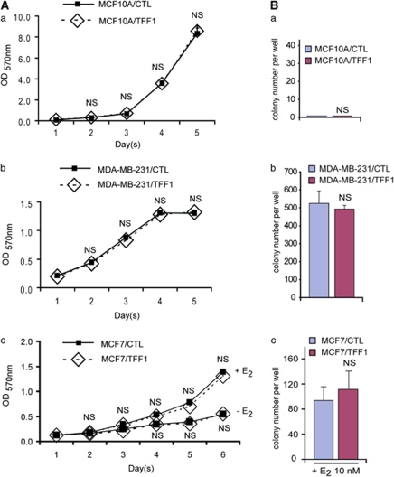
Although trefoil factor 1 (TFF1; previously named pS2) is abnormally expressed in about 50% of human breast tumors, its physiopathological role in this disease has been poorly studied. Moreover, controversial data have been reported. TFF1 function in the mammary gland therefore needs to be clarified. In this study, using retroviral vectors, we performed TFF1 gain- or loss-of-function experiments in four human mammary epithelial cell lines: normal immortalized TFF1-negative MCF10A, malignant TFF1-negative MDA-MB-231 and malignant TFF1-positive MCF7 and ZR75.1. The expression of TFF1 stimulated the migration and invasion in the four cell lines. Forced TFF1 expression in MCF10A, MDA-MB-231 and MCF7 cells did not modify anchorage-dependent or -independent cell proliferation. By contrast, TFF1 knockdown in MCF7 enhanced soft-agar colony formation. This increased oncogenic potential of MCF7 cells in the absence of TFF1 was confirmed in vivo in nude mice. Moreover, chemically induced tumorigenesis in TFF1-deficient (TFF1-KO) mice led to higher tumor incidence in the mammary gland and larger tumor size compared with wild-type mice. Similarly, tumor development was increased in the TFF1-KO ovary and lung. Collectively, our results clearly show that TFF1 does not exhibit oncogenic properties, but rather reduces tumor development. This beneficial function of TFF1 is in agreement with many clinical studies reporting a better outcome for patients with TFF1-positive breast primary tumors.


Figures

Figure 1
Figure 1
Induction of constitutive TFF1 expression in MCF10A, MDA-MB-231 and MCF7 breast epithelial cells. (a) pQCXIP and pQCXIP hTFF1 retroviral vectors. (b) Total proteins from whole-cell lysates (Cell Extr) and conditioned medium (Cond Med) from MCF10A/CTL and MCF10A/TFF1 cells were separated by 10–20% gradient SDS–polyacrylamide gel electrophoresis and subjected to TFF1 immunoblot analysis using the p28O2 antibody. MCF10A/TFF1 cells strongly expressed and secreted TFF1, whereas MCF10A/CTL cells were completely TFF1 negative. (c) Western blot showed that MDA-MB-231/TFF1 cells expressed and secreted TFF1, whereas MDA-MB-231/CTL cells remained TFF1 negative. (d) Western blot analysis of MCF7/CTL and MCF7/TFF1 cells shows that TFF1 is overexpressed regardless of E2 status. Note that, in the presence of E2, endogenous TFF1 is strongly expressed and secreted by MCF7/CTL cells. β-Actin, tubulin and Cath-D served as loading controls.
Figure 2
Figure 2
Impact of constitutive TFF1 on MCF10A, MDA-MB-231 and MCF7 cell migration and invasion. (A) Crystal violet-stained migrated cells present at the lower face of the Transwell membranes were counted with an inverted microscope; (a) increased MCF10A/TFF1 migration compared with MCF10A/CTL (P<0.001); (b) similar results for MDA-MB-231/TFF1 versus MDA-MB-231/CTL cells (P<0.01); (c) similar results for MCF7/TFF1 versus MCF7/CTL cells (P<0.001); no effect was shown for MCF7/TFF1 cells in the presence of E2. (B) Invasive cells through matrigel-coated membranes were analyzed as in A. TFF1 expression increases invasion, regardless of the cell tested (a and c, P<0.001; b, P<0.05). No effect was shown for MCF7/TFF1 cells in the presence of E2. Histograms: mean±s.e. of triplicates. NS, not significant.
Figure 3
Figure 3
Impact of constitutive TFF1 on MCF10A, MDA-MB-231 and MCF7 cell proliferation and colony formation in soft agar. (A) cell proliferation: MCF10A/CTL and MCF10A/TFF1 (a), MDA-MB-231/CTL and MDA-MB-231/TFF1 (b) and MCF7/CTL and MCF7/TFF1 cells (c) were cultured for 5 days in culture medium containing 1% fetal calf serum, and counted using the MTT method. No significant differences (NS) were observed regardless of TFF1 status. Note that MCF7/TFF1 and MCF7/CTL cell proliferation were low in the absence of E2. (B) Cells were agar embedded. After 18 days, colonies were stained (crystal violet) and counted. (a) MCF10A/TFF1 and MCF10A/CTL did not form colonies. (b) MDA-MB-231/TFF1 and MDA-MB-231/CTL cells gave rise to colonies of similar number (NS). (c) In the absence of E2, MCF7/TFF1 and MCF7/CTL colony numbers were very low. However, in the presence of E2 (10 n), the number of colonies increased in both MCF7/TFF1 and MCF7/CTL in a similar manner (NS). Histograms: mean±s.e. of triplicates.
Figure 4
Figure 4
Impact of forced TFF1 expression on MCF7 cell tumorigenicity in nude mice. (a) Tumor occurrence after subcutaneous injection in nude mice of 106 cells. Curves showed that MCF7/CTL and MCF7/TFF1 did not develop tumors in the absence of E2. In the presence of E2, both cell lines developed tumors in a similar manner (NS). (b) Western blot showed that MCF7/CTL tumor cells expressed TFF1 (endogenous TFF1), as well as MCF7/TFF1 (endogenous TFF1 plus pQCXIP TFF1). (c) Tumor immunohistochemistry confirmed TFF1 expression (brown). Bars: 30 μm.
Figure 5
Figure 5
Knockdown of TFF1 expression in MCF7 and ZR75.1 breast epithelial cells. (a) TFF1 messenger RNA complete sequence; the ATG start site and the TAG stop codon are in bold, and the coding sequence (CDS) in yellow. Putative TFF1 shRNA sequences are underlined as indicated. (b) pLMP shScr and pLMP shTFF1 retroviral vectors. (c) Western blot analysis of cell extracts and conditioned medium showed that endogenous TFF1 protein levels were higher in MCF7/shScr than in all MCF7/shTFF1s cells. shTFF1#1 and shTFF1#4 were the most efficient in knocking TFF1 in MCF7 cells. (d) Similarly, ZR75.1/shTFF1#1 and ZR75.1/shTFF1#4 showed a dramatic TFF1 decrease compared with ZR75.1/shScr. Tubulin and Cath-D are loading controls.
Figure 6
Figure 6
Impact of TFF1 knockdown on MCF7 and ZR75.1 cell migration and invasion. (A) Number of crystal violet-stained migrated cells present at the lower face of the Transwell membranes; (a) decreased MCF7/shTFF1#1 and MCF7/shTFF1#4 cell migration compared with MCF7/shScr (P<0.001); (b) similar results for ZR75.1/shTFF1#1 and ZR75.1/shTFF1#4 compared with ZR75.1/shScr (P<0.001). (B) Number of invasive cells through matrigel-coated membranes; (a) decreased MCF7/shTFF1#1 and MCF7/shTFF1#4 cell invasion compared with MCF7/shScr (P<0.001 and P<0.01, respectively); (b) similar results for ZR75.1/shTFF1#1 and ZR75.1/shTFF1#4 compared with ZR75.1/shScr (P<0.001). Histograms: mean±s.e. of triplicates.
Figure 7
Figure 7
Impact of TFF1 knockdown on MCF7 cell proliferation and colony formation in soft agar. (a) Using the MTT method, no differences (NS) were observed between MCF7/shScr, MCF7/shTFF1#1 and MCF7/shTFF1#4 cell proliferation. (b) MCF7/shTFF1#1 and MCF7/shTFF1#4 developed more colonies in soft agar than MCF7/shScr cells (P<0.001 and P<0.01, respectively). Histograms: mean±s.e. of triplicates.
Figure 8
Figure 8
Impact of TFF1 knockdown on MCF7 cell tumorigenicity in nude mice. (a) Tumor occurrence after subcutaneous injection in nude mice of 2.5 × 10⁵ cells in the presence of E2. Curves showed that MCF7/shTFF1#1 tumors appeared earlier, and that their incidence was higher than for MCF7/shScr tumors. (b) TFF1 tumor immunohistochemistry showed that MCF7/shScr tumor cells strongly expressed TFF1, whereas only a few MCF7/shTFF1#1 tumor cells were TFF1 positive (brown). Bars: 30 microns.

References

    1. Amiry N, Kong X, Muniraj N, Kannan N, Grandison PM, Lin J, et al. Trefoil factor-1 (TFF1) enhances oncogenicity of mammary carcinoma cells. Endocrinology. 2009;150:4473–4483. - PubMed
    1. Andrew DJ, Ewald AJ. Morphogenesis of epithelial tubes: Insights into tube formation, elongation, and elaboration. Dev Biol. 2010;341:34–55. - PMC - PubMed
    1. Bossenmeyer-Pourie C, Kannan R, Ribieras S, Wendling C, Stoll I, Thim L, et al. The trefoil factor 1 participates in gastrointestinal cell differentiation by delaying G1-S phase transition and reducing apoptosis. J Cell Biol. 2002;157:761–770. - PMC - PubMed
    1. Buron N, Guery L, Creuzot-Garcher C, Lafontaine PO, Bron A, Rio MC, et al. Trefoil factor TFF1-induced protection of conjunctival cells from apoptosis at premitochondrial and postmitochondrial levels. Invest Ophthalmol Vis Sci. 2008;49:3790–3798. - PubMed
    1. Capony F, Rougeot C, Montcourrier P, Cavailles V, Salazar G, Rochefort H. Increased secretion, altered processing, and glycosylation of pro-cathepsin D in human mammary cancer cells. Cancer Res. 1989;49:3904–3909. - PubMed

Publication types

MeSH terms