. 2011 Feb 16;6(2):e17103.
doi: 10.1371/journal.pone.0017103.

Rituximab therapy reduces organ-specific T cell responses and ameliorates experimental autoimmune encephalomyelitis

Affiliations

Rituximab therapy reduces organ-specific T cell responses and ameliorates experimental autoimmune encephalomyelitis

Nancy L Monson et al. PLoS One. .

Abstract

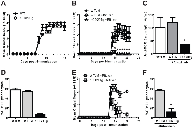
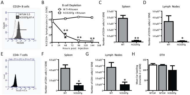
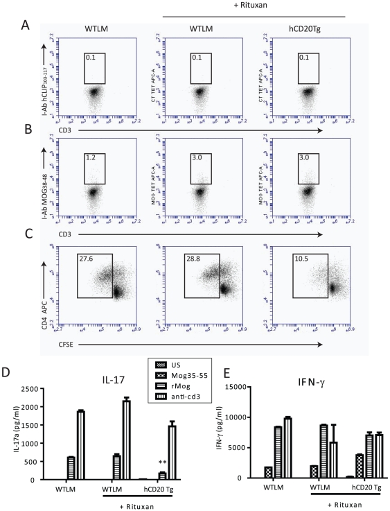
Recent clinical trials have established B cell depletion by the anti-CD20 chimeric antibody Rituximab as a beneficial therapy for patients with relapsing-remitting multiple sclerosis (MS). The impact of Rituximab on T cell responses remains largely unexplored. In the experimental autoimmune encephalomyelitis (EAE) model of MS in mice that express human CD20, Rituximab administration rapidly depleted peripheral B cells and strongly reduced EAE severity. B cell depletion was also associated with diminished Delayed Type Hypersensitivity (DTH) and a reduction in T cell proliferation and IL-17 production during recall immune response experiments. While Rituximab is not considered a broad immunosuppressant, our results indicate a role for B cells as a therapeutic cellular target in regulating encephalitogenic T cell responses in specific tissues.


Conflict of interest statement

Competing Interests: The authors have declared that no competing interests exist.

Figures

Figure 1
Figure 1. Disruption of EAE pathogenesis by B cell depletion.
Panel A. EAE severity is similar in WTLM and hCD20Tg mice. EAE onset and severity were monitored using a 5-point scale on WTLM and hCD20Tg mice immunized with MOG1–125. These results are representative of at least two independent experiments. Panels B–D. Rituximab administration prevents the induction of EAE. WTLM or hCD20Tg mice were either left untreated or were injected with 100 µg Rituximab daily for three days (Day -3,-2,-1). On Day 0, EAE was induced by immunization with MOG1–125. Panel B. Disease course of WTLM and hCD20Tg mice, EAE onset and severity was monitored using a 5-point scale. Shown are the mean clinical score +/− SEM. Panel C. B cell depletion results in reduced levels of anti-MOG IgG in the serum. Serum was harvested on day 21 post-immunization and MOG-specific IgG levels were determined by ELISA. Results shown are the mean IgG concentration +/− SEM. Asterix indicates significant decrease as compared to Rituximab-treated WTLM mice. Panel D. Rituximab administration results in rapid depletion of B cells in the peripheral blood. Blood was taken from WTLM or hCD20Tg mice 3 days following the final dose of Rituximab (day 2 post-immunization). B cells were identified by flow cytometry using gates to identify lymphocytes and CD19 expressing cells. Results shown are the mean percentages of CD19+ B cells +/−SEM (*, p<0.01). Panels E/F. Treatment with Rituximab reduces EAE severity. EAE was initiated in WTLM and hCD20Tg mice on Day 0. Upon the appearance of clinical signs of EAE, Rituximab (100 µg) was administered daily for three treatments. Panel E. Disease course of WTLM and hCD20Tg mice, EAE onset and severity was monitored using a 5-point scale. Shown are the mean clinical score +/− SEM. Panel F. B cell depletion in peripheral blood on day 20. Significant differences were determined using an unpaired t-test (*, p<0.05; **, p<0.01). These results are representative of at least two independent experiments with Rituximab and two experiments using the 1F5 anti-human CD20 mAb (data not shown).
Figure 2
Figure 2. B and T cell dynamics following Rituximab treatment.
Panels A and E. Expression of human CD20 by hCD20Tg B cells and hCD20Tg T cells. Splenocytes from WTLM and hCD20Tg mice were stained with antibodies to human CD20, CD4 and CD19 and flow cytometry performed. Results shown are gated on CD19+CD4− events to identify B cells or gated on CD19−CD4+ events to identify T cells. Panels B/C/D/F/G. WTLM and hCD20Tg mice received three daily injections of Rituximab (100 µg) beginning on day 0. At 144 hours after Rituximab treatment was initiated, tissues were harvested for flow cytometry analysis. Panel B. Peripheral B cells are rapidly depleted following Rituximab treatment. Panel C. Splenic B cells are depleted following Rituximab treatment. Panel D. B cells in the LN (Axilary, Brachial and Inguinal) are depleted following Rituximab treatment. Panel E. CD4 T cells do not express human CD20. Panel F. Splenic CD4 T cells are reduced following Rituximab treatment. Panel G. CD4 T cells in the LN (Axilary, Brachial and Inguinal) are reduced following Rituximab treatment. Panel H. Rituximab administration does not prevent priming of inflammatory T-effector cells. WTLM and hCD20Tg mice were treated with Rituximab daily for 3 days (Day -3,-2,-1), followed by immunization with MOG1–125 on Day 0. On day 10 post-immunization, DTH responses were elicited by subcutaneous injection of MOG1–125 (10 µg) in the ear. The net ear swelling responses were determined at 24 hours. Results shown indicate the mean ear swelling in mmX10E-3 (background subtracted) +/− SEM. Significant differences were detected by unpaired t-test (*, p<0.05; **, p<0.01). These results are representative of at least two independent experiments.
Figure 3
Figure 3. Rituximab administration alters MOG-specific recall responses.
Panels A–E. WTLM and hCD20Tg mice were treated with Rituximab daily for 3 days (Day -3,-2,-1) followed by immunization with MOG1–125 on Day 0. On day 20 post-immunization, bulk draining lymph node cells (LNC) were isolated and recall response determined. Panels A and B. Identification of MOG-reactive T cells by tetramer staining. Bulk LNC were cultured for 3 days in the presence of MOG1–125 prior to labeling with antibodies to CD3, CD4 and I-Ab tetramers to either human (A) CLIP103–117 or (B) MOG38–48. Numbers above boxes indicate percentages of T cells in the tetramer positive gate. Panel C. Secondary T cell proliferative responses were determined by CFSE dilution assay. LNC were labeled with CFSE and placed in culture with 20 µg/ml MOG1–125 and proliferation determined by flow cytometry on day 6 of culture. Results shown are gated on CD4+ events. Numbers indicate the percentage of total cells that diluted CFSE from WTLM and hCD20Tg mice. Panels D and E. 48-hour supernatants from the Panel C experiments were examined for the presence of IL-17 (D) or IFNγ (E) by ELISA. Asterices indicate a significant decrease in IL-17 production (p<0.05). Results are representative of at least 2 independent experiments.

References

    1. Bauman TM, Kasper LH. Novel approaches and cutting edge immunotherapies in multiple sclerosis. Front Biosci. 2004;9:2302–2322. - PubMed
    1. Menge T, Weber MS, Hemmer B, Kieseier BC, von Budingen HC, et al. Disease-modifying agents for multiple sclerosis: recent advances and future prospects. Drugs. 2008;68:2445–2468. - PubMed
    1. Grillo-Lopez AJ, Hedrick E, Rashford M, Benyunes M. Rituximab: ongoing and future clinical developmemt. Semin Oncol. 2002;29:105–112. - PubMed
    1. Stuve O, Cepok S, Elias B, Saleh A, Hartung HP, Hemmer B, et al. Arch Neurol In Press; 2005. Clinical Stabilization and Effective B cell Depletion in the Cerebrospinal Fluid and Peripheral Blood in a Patient with Fulminant Relapsing Remitting Multiple Sclerosis. - PubMed
    1. Hauser SL, Waubant E, Arnold DL, Vollmer T, Antel J, et al. B-cell depletion with Rituximab in relapsing-remitting multiple sclerosis. N Engl J Med. 2008;358:676–688. - PubMed

Publication types

MeSH terms