. 2011 Feb 16;6(2):e17116.
doi: 10.1371/journal.pone.0017116.

Sex hormones in autism: androgens and estrogens differentially and reciprocally regulate RORA, a novel candidate gene for autism

Affiliations

Sex hormones in autism: androgens and estrogens differentially and reciprocally regulate RORA, a novel candidate gene for autism

Tewarit Sarachana et al. PLoS One. .

Abstract

Autism, a pervasive neurodevelopmental disorder manifested by deficits in social behavior and interpersonal communication, and by stereotyped, repetitive behaviors, is inexplicably biased towards males by a ratio of ∼4∶1, with no clear understanding of whether or how the sex hormones may play a role in autism susceptibility. Here, we show that male and female hormones differentially regulate the expression of a novel autism candidate gene, retinoic acid-related orphan receptor-alpha (RORA) in a neuronal cell line, SH-SY5Y. In addition, we demonstrate that RORA transcriptionally regulates aromatase, an enzyme that converts testosterone to estrogen. We further show that aromatase protein is significantly reduced in the frontal cortex of autistic subjects relative to sex- and age-matched controls, and is strongly correlated with RORA protein levels in the brain. These results indicate that RORA has the potential to be under both negative and positive feedback regulation by male and female hormones, respectively, through one of its transcriptional targets, aromatase, and further suggest a mechanism for introducing sex bias in autism.


Conflict of interest statement

Competing Interests: The authors have declared that no competing interests exist.

Figures

Figure 1
Figure 1. Effect of sex hormones on RORA expression in SH-SY5Y cells.
(A) Dose-response to estradiol. The cells were treated with 0.1, 1, or 10 nM 17β-estradiol (E2) for 2 hrs and RORA expression was measured by qRT-PCR analyses (n = 3 per treatment group). (B) Time-course of response to estradiol. The cells were treated with 1 nM 17β-estradiol and qRT-PCR analyses (n = 3 per time point) were conducted to determine RORA expression at different times after hormone addition. (C) Dose-response to DHT. The cells were treated with 0.1, 1, or 10 nM DHT for 2 hrs and RORA expression was measured by qRT-PCR analyses (n = 3 per treatment group). (D) Time-course of response to DHT. The cells were treated with 10 nM DHT and qRT-PCR analyses (n = 3 per time point) were conducted to determine RORA expression at different times after hormone addition. Error bars indicate SEM (*P<0.05, **P<0.01, ***P<0.005, versus mock-treatment control).
Figure 2
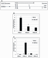
Figure 2. RORA is a potential transcriptional target of both AR and ER.
(A) Schematic diagram showing the upstream region of the RORA gene (edited from the UCSC Genome Browser). Potential AR and ER binding sites are labeled (ARbs  =  AR potential binding site, ERbs  =  ER potential binding site). (B) Chromatin Immunoprecipitation followed by qPCR (ChIP-qPCR) analyses of AR potential binding sites on the RORA gene promoter region. Sonicated chromatin from SH-SY5Y cells treated with 1 nM DHT for 2 hrs was immunoprecipitated (IP) using anti-AR or IgG antibody. Parallel samples were mock-treated with the hormone delivery vehicle, ethanol. The enrichment of each AR binding site in anti-AR-IP DNA was determined by qPCR analyses (n = 3), and normalized to the enrichment in IgG-IP DNA (control). Error bars indicate SEM (*P<0.01 versus mock-treatment control). (C) ChIP-qPCR analyses of ERα potential binding sites on the RORA gene promoter region. Sonicated chromatin from SH-SY5Y cells treated with 1 nM 17β-estradiol (or vehicle) for 2 hrs was immunoprecipitated using anti-ERα or control IgG antibody. The enrichment of each ERα binding site in anti-ERα-IP DNA was determined by qPCR analyses (n = 3), and normalized to the enrichment in IgG-IP DNA. Error bars indicate SEM (#Undetectable amount of ERα binding sites in the mock-treatment control).
Figure 3
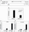
Figure 3. Aromatase is a potential transcriptional target of RORA.
(A) Schematic diagram showing the upstream region of the aromatase gene (edited from the UCSC Genome Browser). Potential RORA binding sites are labeled (RORAbs  =  RORA potential binding sites). Because sites II and III are so close to each other, a single pair of primers was designed to cover both sites. (B) ChIP-qPCR analyses of potential binding sites for RORA on the aromatase gene promoter region. Sonicated chromatin from SH-SY5Y cells was immunoprecipitated using anti-RORA or control IgG antibody. The enrichment of each RORA binding site in anti-RORA-IP DNA was determined by qPCR analyses (n = 3). Error bars indicate SEM (*P<0.05, **P<0.001, versus the enrichment in IgG-IP DNA). Aromatase expression is enhanced by RORA overexpression. SH-SY5Y cells were transfected with 1.25 µg or 2.50 µg pSG5.HA-RORA plasmid or empty plasmid for 24 hrs, and qRT-PCR analyses (n = 3) were conducted to measure RORA and aromatase expression (Mock  =  mock-treatment control, Neg  =  empty plasmid, RORA  =  pSG5.HA-RORA plasmid). (C) Relative quantity of RORA transcript levels in RORA-transfected cells which is increased by more than a factor of 105 relative to the controls. (**P<0.001 versus the empty plasmid control) (D) Relative quantity of aromatase transcript levels in RORA-transfected cells. Error bars indicate SEM (*P<0.05 versus the empty plasmid control).
Figure 4
Figure 4. RORA and aromatase proteins are decreased in postmortem brain tissues from autistic subjects.
A tissue array containing postmortem frontal cortex specimen from 12 autistic individuals and 22 age- and sex-matched controls was co-immunolabeled for aromatase (red), RORA (blue), and MAP2, and counterstained with DAPI to reveal nuclei (see Fig. S1 for example of quadruple staining). (A) Representative images from confocal immunofluorescence analyses of the frontal cortex tissue arrays. Arrows mark neurons in the frontal cortex. Scale bars, 20 µm. F: female; M: male; #'s indicate the respective ages of the subjects from whom the brain tissues were obtained. (B) Quantitative analyses of confocal fluorescence images of RORA, aromatase, and DAPI. Frontal cortex neurons expressing MAP2 protein were selected according to its green immunofluorescence (shown in Fig. S1), and fluorescence signal intensity for RORA, aromatase (ARO), and DAPI, in the neurons were extracted using image analysis software and normalized by background subtraction. For each sample, ∼40–50 neurons were selected for quantification of RORA and aromatase fluorescence in a “sample blind” fashion such that the identities of the samples were unknown to the person performing the analyses. Error bars indicate SEM (*P<0.05 versus the control samples). (C) Correlation analysis of RORA and aromatase protein expression in the frontal cortex neurons.
Figure 5
Figure 5. A model for reciprocal hormonal effects on RORA.
The schematic illustrates a mechanism through which the observed reduction in RORA in autistic brain may lead to increased testosterone levels through downregulation of aromatase. Through AR, testosterone negatively modulates RORA, whereas estrogen upregulates RORA through ER.

References

    1. American Psychological Association. Washington, DC: American Psychological Association; 1994. Diagnostic and statistical manual of mental disorders.
    1. Volkmar FR, Klin A, Siegel B, Szatmari P, Lord C, et al. Field trial for autistic disorder in DSM-IV. Am J Psychiatry. 1994;151(9):1361–7. - PubMed
    1. Baron-Cohen S, Knickmeyer RC, Belmonte MK. Sex differences in the brain: Implications for explaining autism. Science. 2005;310(5749):819–23. - PubMed
    1. Auyeung B, Taylor K, Hackett G, Baron-Cohen S. Foetal testosterone and autistic traits in 18 to 24-month-old children. Molecular Autism. 2010;1(1):11. - PMC - PubMed
    1. Auyeung B, Baron-Cohen S, Ashwin E, Knickmeyer R, Taylor K, et al. Fetal testosterone and autistic traits. Br J Psychol. 2009;100(1):1–22. - PubMed

Publication types

MeSH terms

LinkOut - more resources