. 2011 Jul;12(2):63-76.
doi: 10.1007/s10969-011-9102-6. Epub 2011 Feb 26.

Leveraging structure determination with fragment screening for infectious disease drug targets: MECP synthase from Burkholderia pseudomallei

Affiliations

Leveraging structure determination with fragment screening for infectious disease drug targets: MECP synthase from Burkholderia pseudomallei

Darren W Begley et al. J Struct Funct Genomics. 2011 Jul.

Abstract

As part of the Seattle Structural Genomics Center for Infectious Disease, we seek to enhance structural genomics with ligand-bound structure data which can serve as a blueprint for structure-based drug design. We have adapted fragment-based screening methods to our structural genomics pipeline to generate multiple ligand-bound structures of high priority drug targets from pathogenic organisms. In this study, we report fragment screening methods and structure determination results for 2C-methyl-D-erythritol-2,4-cyclo-diphosphate (MECP) synthase from Burkholderia pseudomallei, the gram-negative bacterium which causes melioidosis. Screening by nuclear magnetic resonance spectroscopy as well as crystal soaking followed by X-ray diffraction led to the identification of several small molecules which bind this enzyme in a critical metabolic pathway. A series of complex structures obtained with screening hits reveal distinct binding pockets and a range of small molecules which form complexes with the target. Additional soaks with these compounds further demonstrate a subset of fragments to only bind the protein when present in specific combinations. This ensemble of fragment-bound complexes illuminates several characteristics of MECP synthase, including a previously unknown binding surface external to the catalytic active site. These ligand-bound structures now serve to guide medicinal chemists and structural biologists in rational design of novel inhibitors for this enzyme.


Figures

Fig. 1
Fig. 1
a STD-NMR and 2D NOESY spectra with b zoomed-in region for a sample containing BpIspF, cytidine and 34 small molecule fragments from primary screening. Peaks visible in the STD spectrum (purple) and positive crosspeaks (red) generated by negative NOE signals are indicative of small molecule binders. c Chemical structures for all compounds in this mixture; binders are boxed and labeled, including FOL535 which was previously observed to bind BpIspF by X-ray crystallography (PDB: 3K14). Figure generated using iNMR software (http://www.inmr.net)
Fig. 2
Fig. 2
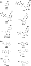
Chemical structures and PDB codes for a cytosine pocket, b zinc-site, and c external site BpIspF-binding fragments
Fig. 3
Fig. 3
Small molecules which bind BpIspF fall into three distinct categories: a cytidine pocket binders, including cytosine (yellow), cytidine (cyan), 5′-iodo-cytidine (green), CMP (navy), CDP (magenta) and CTP (white); b zinc-site binders FOL535 (magenta), FOL717 (navy), FOL8395 (cyan) and FOL955 (white); and c external site binders FOL694 (magenta) and FOL795 (cyan). A single protein crystal structure (PDB ID: 3P10) is depicted for clarity. Key interactions illustrated in a between cytidine, D48 of one monomer, and A102, P105 and A108 of the opposite monomer (black dashes). Cytidine and FOL955 (white) are illustrated in the active site for c. Figure generated using PyMol [57]
Fig. 4
Fig. 4
Structure ensemble of fragments bound to 2C-methyl-D-erythritol-2,4-cyclo-diphosphate synthase from Burkholderia pseudomallei (PDB IDs 3IEQ, 3IEW, 3IKE, 3IKF, 3JVH, 3K14, 3K2X, 3MBM, 3P0Z, 3P10 and 3QHD). The holoenzyme possesses three active sites, located in a solvent-exposed groove along each monomer–monomer interface (green, cyan, magenta). Each active site also contains a catalytic zinc ion (yellow spheres). Molecules of tris, glycerol, and acetate were observed to bind in the center of the trimeric protein, but no FOL compounds were found in this site. For clarity, a single protein crystal structure (PDB ID:3P10) is viewed along the threefold trimer axis. Figure generated using PyMol [57]

References

    1. Myler PJ, Stacy R, Stewart L, Staker BL, Van Voorhis WC, Varani G, Buchko GW. The Seattle Structural Genomics Center for Infectious Disease (SSGCID) Infect Disord Drug Targets. 2009;9(5):493–506. doi: 10.2174/187152609789105687. - DOI - PMC - PubMed
    1. Van Voorhis WC, Hol WG, Myler PJ, Stewart LJ (2009) The role of medical structural genomics in discovering new drugs for infectious diseases. PLoS Comput Biol 5(10):e1000530 - PMC - PubMed
    1. Davies DR, Mamat B, Magnusson OT, et al. Discovery of leukotriene A4 hydrolase inhibitors using metabolomics biased fragment crystallography. J Med Chem. 2009;52(15):4694–4715. doi: 10.1021/jm900259h. - DOI - PMC - PubMed
    1. Hartshorn MJ, Murray CW, Cleasby A, Frederickson M, Tickle IJ, Jhoti H. Fragment-based lead discovery using X-ray crystallography. J Med Chem. 2005;48(2):403–413. doi: 10.1021/jm0495778. - DOI - PubMed
    1. Brough PA, Barril X, Borgognoni J, et al. Combining hit identification strategies: fragment-based and in silico approaches to orally active 2-aminothieno[2, 3-d]pyrimidine inhibitors of the Hsp90 molecular chaperone. J Med Chem. 2009;52(15):4794–4809. doi: 10.1021/jm900357y. - DOI - PubMed

Publication types