. 2011 Jul;12(2):83-95.
doi: 10.1007/s10969-011-9101-7. Epub 2011 Feb 27.

SAD phasing using iodide ions in a high-throughput structural genomics environment

Affiliations

SAD phasing using iodide ions in a high-throughput structural genomics environment

Jan Abendroth et al. J Struct Funct Genomics. 2011 Jul.

Abstract

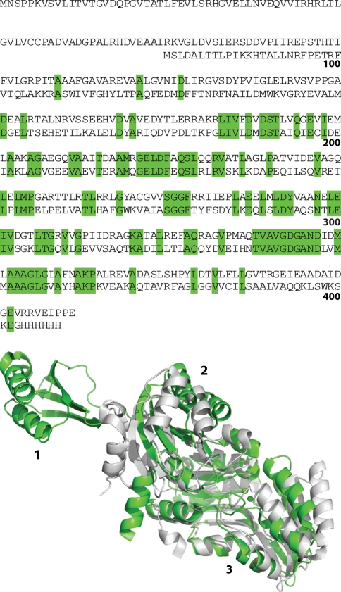
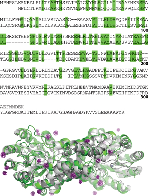
The Seattle Structural Genomics Center for Infectious Disease (SSGCID) focuses on the structure elucidation of potential drug targets from class A, B, and C infectious disease organisms. Many SSGCID targets are selected because they have homologs in other organisms that are validated drug targets with known structures. Thus, many SSGCID targets are expected to be solved by molecular replacement (MR), and reflective of this, all proteins are expressed in native form. However, many community request targets do not have homologs with known structures and not all internally selected targets readily solve by MR, necessitating experimental phase determination. We have adopted the use of iodide ion soaks and single wavelength anomalous dispersion (SAD) experiments as our primary method for de novo phasing. This method uses existing native crystals and in house data collection, resulting in rapid, low cost structure determination. Iodide ions are non-toxic and soluble at molar concentrations, facilitating binding at numerous hydrophobic or positively charged sites. We have used this technique across a wide range of crystallization conditions with successful structure determination in 16 of 17 cases within the first year of use (94% success rate). Here we present a general overview of this method as well as several examples including SAD phasing of proteins with novel folds and the combined use of SAD and MR for targets with weak MR solutions. These cases highlight the straightforward and powerful method of iodide ion SAD phasing in a high-throughput structural genomics environment.


Figures

Fig. 1
Fig. 1
Anomalous scattering factors for iodide and selenium across the energy range used in macromolecular crystallography. The image was generated using the University of Washington X-ray Anomalous Scattering Server developed and maintained by Ethan A. Merritt (http://skuld.bmsc.washington.edu/scatter/) which is based on an earlier publication [59]
Fig. 2
Fig. 2
Crystal structures determined by SSGCID using iodide ion soaks and SAD phasing
Fig. 3
Fig. 3
Types of iodide ion binding sites. a Arginine-iodide ion interaction network on the surface of MysmA.17112.a (PDB ID 3OL3), a putative uncharacterized protein from Mycobacterium smegmatis and an ortholog of community request protein MytuD.17112.a, Rv0543c from Mycobacterium tuberculosis (PDB ID 2KVC [60]) b Iodide ions binding along an α-helix of CoimA.00345.a, a putative fructose-1,6-bisphosphate aldolase from Coccidioides immits (PDB ID 3PM6). Iodide ion IA forms a possible anion–cation interaction with His21, while forming an interaction with the side chain hydroxyl of Thr20 (3.3 Å), an amide interaction with Thr16 and a hydrophobic interaction with Phe17. Iodide ion IB forms hydrophobic interactions with the side chains of Met298 and Val14, while packing off the amide of Pro13-Val14. c Iodide ion binding to the periplasmic domain of the risS pH sensor histidine kinase from Burkholderia pseudomallei (BupsA.00863.i, a community request target, PDB ID 3LR0). The iodide ion forms an interaction with the backbone amide nitrogen of Asp122 (3.5 Å) while forming another interaction with the side chain of Ser119 (3.4 Å) and packing against two β-sheets. d Iodide ion binding off reduced flavin adenine dinucleotide (FADH2) in the crystal structure of an acyl-CoA dehydrogenase from M. thermoresistibile (MythA.00185.b, PDB ID 3PFD). An unbiased |F o| − |F c| map calculated from a model lacking the cofactor is shown in green mesh contoured at 3.0 σ. For each panel iodide ions are shown as magenta spheres and an anomalous difference Fourier map is shown in magenta mesh contoured at 5.0 σ
Fig. 4
Fig. 4
Overlay of NMR solution structures of BolA-like proteins from M. musculus (gray, PDB ID 1V9J [42]), P. falciparum (magenta, Buchko, G.W. et al. unpublished) and the crystal structure of a BolA-like protein from B. bovis solved by iodide ion SAD (green, PDB ID 3O2E). Iodide ions are shown as green spheres. For simplicity only the ordered regions are shown
Fig. 5
Fig. 5
Sequence alignment and crystal structures of phosphoserine phosphatase SerB from Vibrio cholera (bottom sequence, gray ribbons, PDB ID 3N28, Patskovsky, Y. et al. unpublished) and Mycobacterium avium solved by iodide ion SAD (top sequence, green ribbons, PDB ID 3P96). For simplicity, only one monomer of the biological dimer is shown in each case. Domains 1, 2, and 3 of Ma SerB correspond to residues 1–85, 97–175, and 182–400, respectively
Fig. 6
Fig. 6
Sequence alignment and crystal structures of fructose-1,6-bisphosphate aldolases from Giardia lamblia (bottom sequence, gray ribbons, PDB ID 2ISV [48]) and Coccidioides immitis solved by combined iodide ion SAD and MR (top sequence, green ribbons, PDB ID 3PM6). Iodide ions are shown as magenta spheres and the catalytic zinc ions are shown as gray spheres. Anomalous difference Fourier maps are shown in magenta mesh contoured at 5.0 σ

References

    1. Myler PJ, Stacy R, Stewart L, Staker BL, Van Voorhis WC, et al. The Seattle Structural Genomics Center for Infectious Disease (SSGCID) Infect Disord Drug Targets. 2009;9:493–506. doi: 10.2174/187152609789105687. - DOI - PMC - PubMed
    1. Van Voorhis WC, Hol WG, Myler PJ, Stewart LJ. The role of medical structural genomics in discovering new drugs for infectious diseases. PLoS Comput Biol. 2009;5:e1000530. doi: 10.1371/journal.pcbi.1000530. - DOI - PMC - PubMed
    1. Anderson WF. Structural genomics and drug discovery for infectious diseases. Infect Disord Drug Targets. 2009;9:507–517. doi: 10.2174/187152609789105713. - DOI - PMC - PubMed
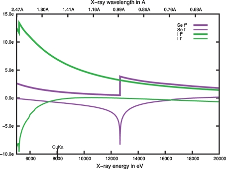
    1. Dauter M, Dauter Z. Phase determination using halide ions. Methods Mol Biol. 2007;364:149–158. - PubMed
    1. Dauter Z, Dauter M, Rajashankar KR. Novel approach to phasing proteins: derivatization by short cryo-soaking with halides. Acta Crystallogr D Biol Crystallogr. 2000;56:232–237. doi: 10.1107/S0907444999016352. - DOI - PubMed

Publication types

MeSH terms

LinkOut - more resources