Review
. 2011 May;22(5):785-801.
doi: 10.1007/s10552-011-9745-4. Epub 2011 Feb 27.

Genomics of the NF-κB signaling pathway: hypothesized role in ovarian cancer

Affiliations
Review

Genomics of the NF-κB signaling pathway: hypothesized role in ovarian cancer

Kristin L White et al. Cancer Causes Control. 2011 May.

Abstract

Objective: We sought to review evidence linking nuclear factor-kappa B (NF-κB) to ovarian cancer and to identify genetic variants involved in NF-κB signaling.

Methods: PubMed was reviewed to inform on ovarian cancer biology and NF-κB signaling and to identify key genes. Public linkage disequilibrium (LD) data were analyzed to identify informative inherited variants (tagSNPs) using ldSelect.

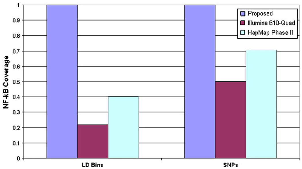
Results: We identified 319 key NF-κB genes including five NF-κB subunits, 167 activating genes, and 55 inhibiting genes. We found that the 1000 Genomes Project was the most informative LD source for most genes (92.8%), and we identified 13,027 LD bins (r (2) ≥ 0.9, minor allele frequency ≥ 0.05) and 1,018 putative-functional variants worthy of investigation. We also report that reliance on a commonly used genome-wide SNP array and genotype imputation with HapMap Phase II data provides data on only 74% of the common inherited NF-κB SNPs of interest.

Conclusions: Compelling evidence suggests that NF-κB plays a critical role in ovarian cancer, yet inherited variation in these genes has not been thoroughly assessed in relation to disease risk or outcome. We present a collection of variants in key genes and suggest creation of a custom genotyping array as an optimal approach.


Figures

Fig. 1
Fig. 1
Key components of NF-κB signaling pathways. Color-coding of molecules: activators = yellow, NF-κB subunits = blue, inhibitors = red, degradators = green, nuclear function = cream. Canonical pathway: Binding of extracellular signaling molecules to receptors leads to recruitment of activation proteins, phosphorylation of IKKβ, activation of the IKK complex (IKKα, IKKβ, and NEMO), and processing of p105 and p100 into functional p50 and p52 by the ubiquitin ligase SCFβ-TrCP complex. Activated IKK complex phosphorylates IκB, marking it for ubiquitination by the SCFβ-TrCP complex and degradation. The main canonical NF-κB dimer, p65/p50, is activated and translocates to the nucleus to initiate gene transcription with the RNA polymerase II preinitiation complex (PIC). Non-canonical pathway: Ligase and receptor coupling leads to NIK and IKKα activation, which initiates processing of p100 into functional p52 by the SCFβ-TrCP complex. The p52/Rel-B dimer translocates to the nucleus to initiate gene expression through the RNA polymerase II PIC
Fig. 2
Fig. 2
TNFα and TCR activation pathways. A TNFα binding to its receptors TNF-R1 and TNF-R2 triggers a coupling between TNF-R1/2 and TRADD via their death domains (DD). TRADD recruits TRAF2, RIPK1, RIPK2, and RIPK3, and TRAF5. Following TNF-R1 endocytosis, TRADD dissociates from the receptor complex and associates with FADD and caspase-8, initiating pro-apoptotic signaling pathways. After TRAF2 auto-poly-ubiquitination, RIPK1 is poly-ubiquitinated, which recruits TAB 1, TAB 2, TAB 3, TAK1, and TRAF6 to the complex. TAB 2 forms a bridge between TRAF6 and TAK1, allowing TAK1 to activate the IKK complex by phosphorylating IKKβ. B Activation of the TCR-CD3 complex induces phosphorylation of ITAMs on TCRζ and CD3. ZAP70 and LCK are recruited, LCK phosphorylates ZAP70, PLCγ1, and the scaffold protein SLP-76, and recruits PKCθ to the complex. PKCθ recruits CARMA1, CARD9, CARD10, CARD14, BCL-10, and MALT1, leading to activation of the IKK complex
Fig. 3
Fig. 3
IL-1 and TLR activation pathways. A Ligand binding to IL-1R1 leads to coupling of IL-1RAPcP1, IL-1RAPcP2, and MYD88 with receptors. MYD88 recruits IRAK-1 and the adaptor proteins Tollip, TRAF6, and IRAK-4 to the receptor complex. After IRAK-1 phosphorylation by IRAK-4, IRAK-1, and TRAF6 dissociate from the receptor complex and associate with TAB 1, TAB 2, TAB 3, and TAK1. Pellino proteins phosphorylate IRAK-1, initiating IRAK-1 and Pellino poly-ubiquitination and degradation, freeing the TRAF6/TAB 1/TAB 2/TAB 3/TAK1 complex. TAB 2 phosphorylates TAK1, which phosphorylates and activates IKKβ. B Toll-like receptors (TLRs) are critical components of the innate immune response as they are conserved pattern recognition receptors (PRRs) that detect conserved pathogen-associated microbial patterns (PAMPs) present on many antigens. There are over ten TLRs that have been identified, all of which recognize a variety of different PAMPs and lead to NF-#x003BA;B activation. TLRs and IL-1R1 have a shared Toll-IL-1R (TIR) domain that mediates interactions with downstream molecules. Due to this shared TIR domain, TLRs initiate a similar signaling cascade as IL-1 involving MYD88, IRAK-1, IRAK-4, TRAF6, TAB 2, and TAK1, but also involve other cascades that include TIRAP, TRIF, TANK, and TBK1
Fig. 4
Fig. 4
Non-canonical activation pathways. Receptor binding of LTβ, BAFF, CD40, and RANK initiate the non-canonical signaling pathway. TRAF proteins and other intracellular target molecules are recruited, eventually leading to the activation of NIK through an unknown mechanism. NIK contains binding domains that interact with TRAF2, TRAF3, TRAF5, and TRAF6, but the specific roles of TRAF and other proteins in the non-canonical pathway is still controversial. After NIK phosphorylation, IKKα phosphorylates p100, marking it for poly-ubiquitination and partial degradation by the SCFβ-TrCP complex and 26S proteasome. Partial degradation of p100 yields functional p52, which most commonly forms heterodimers with Rel-B and translocates to the nucleus to initiate gene transcription
Fig. 5
Fig. 5
Ubiquitination and proteasomal degradation of IκBs. The E3 protein-ubiquitin ligase SCFβ-TrCP complex consists of β-TrCP, an F-box protein with a WD repeat region that serves as the docking subunit for the SCFβ-TrCP complex, the adaptor proteins Skp1 and Cul1, and the RING protein Roc1 that recruits the E2 ubiquitin-conjugating enzyme UbcH5. The precursor proteins p100 and p105 are partially degraded to yield functional p52 and p50, respectively. p105 contains a glucine-rich region (GRR), which separates the functional from inhibitory portions of the protein and is essential for its processing. After being marked for ubiquitination by the SCFβ-TrCP complex, the proteasome degrades p105 and p100 until it hits the GRR region signaling the completion of processing and the functional subunit is released, or the proteasome is unable to properly unfold the GRR region, forcing it to complete processing prematurely and release p50/p65. Additionally, the SCFβ-TrCP complex processes IκB after IKKβ phosphorylates IκBα, IκBβ, and IκBε. β-TrCP recognizes the degradation signal and UbcH5 and the SCFβ-TrCP complex polyubiquitinate lysine residues on IκBα, which marks it for degradation by the 26S proteasome
Fig. 6
Fig. 6
RNA polymerase II transcription complex. Formation of the transcription pre-initiation complex (PIC) begins with the general transcription factor TFII-D, which is composed of the TATA-binding protein TBP and 14 TBP-associated factors (TAFs). The transcription factor, TBP binds to the TATA box in the promoter region and is necessary for completion of the PIC complex. TAF1 and TAF5 appear to be the “organizers” of the complex with TAF1 serving as the scaffold for TAF2, TAF4, TAF5, TAF6, TAF9, TAF11, and TAF12 and branching across two of three lobes in TFII-D. TAF5 binds to TAF1, TAF7, TAF11, TAF12, TAF13, and TBP and contains connections to all three of the TFII-D lobes. Creation of the TFII-D complex recruits TFII-A to bind to both the DNA and TFII-D, stabilizing their interaction. TFII-B binds to TBP and the DNA, serving as a scaffold for RNA polymerase II. TFII-F enters the complex with RNA polymerase II, both of which bind to the TFII-B/TBP complex. After RNA polymerase II is bound, TFII-E and TFII-H enter, completing the PIC and initiating transcription
Fig. 7
Fig. 7
Coverage of inherited variation in 319 NF-κB signaling genes. Coverage calculations are based on tagSNPs in NF-κB genes (r2 ≥ 0.9 and MAF ≥ 0.05) using the most informative source available, including 1000 Genomes Project Low-coverage Pilot data for 92.8% of genes (see “Methods”). LD Bins reflects the proportion of LD bins with at least one tagSNP on the Infinium HD Human610-Quad BeadChip genome-wide SNP panel (Illumina 610-Quad) or imputable based on HapMap Phase II data. SNPs reflect the proportion of SNPs interrogated, either directly or indirectly, via genotyping the included tagSNPs on the Illumina 610-Quad or imputing based on HapMap Phase II data (representing SNPs directly assessed or in the covered LD bins). Results for the Illumina 610-Quad and HapMap Phase II are presented due to use in current ovarian cancer genome-wide association studies (e.g., Song et al. [65])

Similar articles

Cited by

  • Symbiotic prodrugs (SymProDs) dual targeting of NFkappaB and CDK.
    Rana S, Kour S, Sonawane YA, Robb CM, Contreras JI, Kizhake S, Zahid M, Karpf AR, Natarajan A. Rana S, et al. Chem Biol Drug Des. 2020 Aug;96(2):773-784. doi: 10.1111/cbdd.13684. Epub 2020 Apr 22. Chem Biol Drug Des. 2020. PMID: 32237047 Free PMC article.
  • Variation in NF-κB signaling pathways and survival in invasive epithelial ovarian cancer.
    Block MS, Charbonneau B, Vierkant RA, Fogarty Z, Bamlet WR, Pharoah PD; Georgia Chenevix-Trench; for AOCS; /ACS Group; Rossing MA, Cramer D, Pearce CL, Schildkraut J, Menon U, Kjaer SK, Levine DA, Gronwald J, Culver HA, Whittemore AS, Karlan BY, Lambrechts D, Wentzensen N, Kupryjanczyk J, Chang-Claude J, Bandera EV, Hogdall E, Heitz F, Kaye SB, Fasching PA, Campbell I, Goodman MT, Pejovic T, Bean YT, Hays LE, Lurie G, Eccles D, Hein A, Beckmann MW, Ekici AB, Paul J, Brown R, Flanagan JM, Harter P, du Bois A, Schwaab I, Hogdall CK, Lundvall L, Olson SH, Orlow I, Paddock LE, Rudolph A, Eilber U, Dansonka-Mieszkowska A, Rzepecka IK, Ziolkowska-Seta I, Brinton LA, Yang H, Garcia-Closas M, Despierre E, Lambrechts S, Vergote I, Walsh CS, Lester J, Sieh W, McGuire V, Rothstein JH, Ziogas A, Lubiński J, Cybulski C, Menkiszak J, Jensen A, Gayther SA, Ramus SJ, Gentry-Maharaj A, Berchuck A, Wu AH, Pike MC, Van Den Berg D, Terry KL, Vitonis AF, Ramirez SM, Rider DN, Knutson KL, Sellers TA, Phelan CM, Doherty JA, Johnatty SE, deFazio A, Song H, Tyrer J, Kalli KR, Fridley BL, Cunningham JM, Goode EL. Block MS, et al. Cancer Epidemiol Biomarkers Prev. 2014 Jul;23(7):1421-7. doi: 10.1158/1055-9965.EPI-13-0962. Epub 2014 Apr 16. Cancer Epidemiol Biomarkers Prev. 2014. PMID: 24740199 Free PMC article.
  • The Unique Molecular and Cellular Microenvironment of Ovarian Cancer.
    Worzfeld T, Pogge von Strandmann E, Huber M, Adhikary T, Wagner U, Reinartz S, Müller R. Worzfeld T, et al. Front Oncol. 2017 Feb 22;7:24. doi: 10.3389/fonc.2017.00024. eCollection 2017. Front Oncol. 2017. PMID: 28275576 Free PMC article. Review.
  • A High-Fat and High-Carbohydrate Diet Promotes Reminiscent Hallmarks of an Aging Ovary in the Rabbit Model.
    Díaz-Hernández V, Montaño LM, Caldelas I, Marmolejo-Valencia A. Díaz-Hernández V, et al. Biomedicines. 2022 Nov 29;10(12):3068. doi: 10.3390/biomedicines10123068. Biomedicines. 2022. PMID: 36551824 Free PMC article.
  • Pre-diagnostic serum levels of inflammation markers and risk of ovarian cancer in the prostate, lung, colorectal and ovarian cancer (PLCO) screening trial.
    Trabert B, Pinto L, Hartge P, Kemp T, Black A, Sherman ME, Brinton LA, Pfeiffer RM, Shiels MS, Chaturvedi AK, Hildesheim A, Wentzensen N. Trabert B, et al. Gynecol Oncol. 2014 Nov;135(2):297-304. doi: 10.1016/j.ygyno.2014.08.025. Epub 2014 Aug 23. Gynecol Oncol. 2014. PMID: 25158036 Free PMC article.

References

    1. Jemal A, Siegel R, Xu J, Ward E. Cancer statistics, 2010. CA Cancer J Clin. 2010;60(5):277–300. - PubMed
    1. Hoskins WJ, Bundy BN, Thigpen JT, Omura GA. The influence of cytoreductive surgery on recurrence-free interval and survival in small-volume stage III epithelial ovarian cancer: a Gynecologic Oncology Group study. Gynecol Oncol. 1992;47(2):159–166. - PubMed
    1. McGuire V, Jesser CA, Whittemore AS. Survival among U.S. women with invasive epithelial ovarian cancer. Gynecol Oncol. 2002;84(3):399–403. - PubMed
    1. Barnholtz-Sloan JS, Schwartz AG, Qureshi F, Jacques S, Malone J, Munkarah AR. Ovarian cancer: changes in patterns at diagnosis and relative survival over the last three decades. Am J Obstet Gynecol. 2003;189(4):1120–1127. - PubMed
    1. Shih Ie M, Kurman RJ. Ovarian tumorigenesis: a proposed model based on morphological and molecular genetic analysis. Am J Pathol. 2004;164(5):1511–1518. - PMC - PubMed

Publication types