. 2011 Dec;469(12):3371-8.
doi: 10.1007/s11999-011-1820-3.

A reliable radiographic measurement technique for extra-articular scapular fractures

Affiliations

A reliable radiographic measurement technique for extra-articular scapular fractures

Jack Anavian et al. Clin Orthop Relat Res. 2011 Dec.

Abstract

Background: Currently, neither well-defined nor standardized measurement techniques exist for assessing deformity of extra-articular scapular fractures. To properly evaluate these injuries, compare observations across studies, and make clinical decisions, a validated measurement protocol for evaluating scapular fractures is needed.

Questions/purposes: We describe techniques to quantitatively characterize extra-articular scapular fracture deformity; evaluate the reliability of these characterizations in plain film radiographs and CT scans; and determine potential differences in the characterization of the deformity between the two imaging modalities.

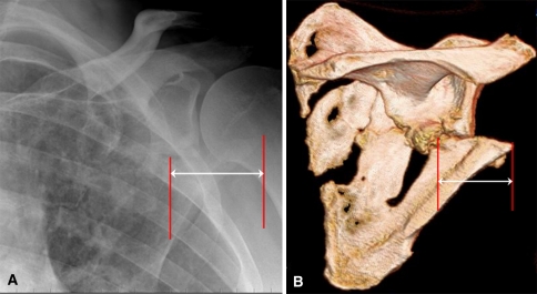
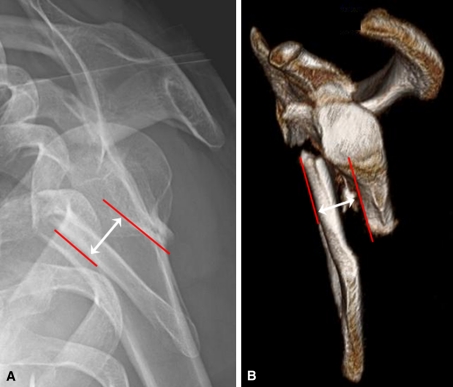
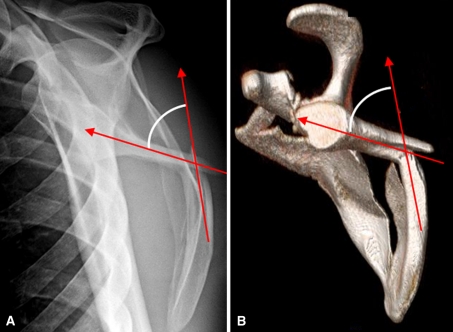
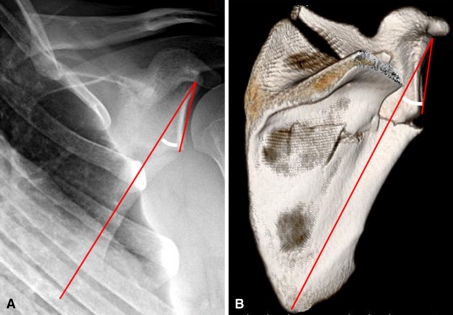
Patients and methods: We evaluated injury radiographs and three-dimensional CT images of 45 patients with extra-articular scapular fracture. Techniques for measuring medial/lateral displacement, angulation, translation, glenopolar angle, and glenoid version were established and utilized in two trials, performed 6 weeks apart, by three observers. We determined descriptive statistics for each measurement parameter.

Results: Interobserver reliability based upon interclass correlation coefficients ranged from 0.36 to 0.76 for radiographs and from 0.48 to 0.87 for three-dimensional CT. Intraobserver reliability using Pearson r coefficient ranged from 0.60 to 0.75 for radiographs and 0.64 to 0.89 for three-dimensional CT. Both individual and pooled measurements for angulation and glenopolar angle were higher on three-dimensional CT versus radiographs.

Conclusions: Our data suggest three-dimensional CT is more reliable than plain radiography in the assessment of scapula fracture displacement. Therefore, we believe this modality should be utilized if fracture deformity warrants surgical consideration and to adequately compare data across studies.

Level of evidence: Level IV, diagnostic study. See Guidelines for Authors for a complete description of levels of evidence.


Figures

Fig. 1A–B
Fig. 1A–B
Measurement of M/L displacement on (A) an AP radiograph and (B) a PA 3D CT scan of the shoulder is demonstrated. M/L = mediolateral; PA = posterior–anterior; 3D = three-dimensional.
Fig. 2A–B
Fig. 2A–B
Measurement of translation on (A) a transcapular Y radiograph and (B) a 3D CT scan is demonstrated. This represents the displacement marked by the distance between the anterior cortex of the proximal fragment and the anterior cortex of the distal fragment. 3D = three-dimensional.
Fig. 3A–B
Fig. 3A–B
Measurement of angulation on (A) a transcapular Y radiograph and (B) a 3D CT scan is demonstrated. A line is drawn through the proximal fragment in parallel with the cortices just proximal to the fracture. A second line is drawn through the distal fragment in parallel with the cortices just distal to the fracture. The angle formed by these two intersecting lines represents the angulation Note, even though the inferior teardrop forms a concave surface over the rib cage, it is the more proximal straight portion of the intramedullary canal that is used for the measurement (a critical distinction in measuring angular deformity so as not to overcall the angulation). 3D = three-dimensional.
Fig. 4A–B
Fig. 4A–B
Measurement of the GPA on (A) a true AP radiograph and (B) a PA 3D CT scan of the shoulder is demonstrated. A line is drawn from the inferior pole of the glenoid fossa up to the superior pole. Another line is drawn from the superior pole of the glenoid fossa down through the inferior-most angle of the scapula body. The angle formed by these two intersecting lines represents the GPA. Normal GPAs range from 30° to 45° [6]. GPA = glenopolar angle; 3D = three-dimensional.
Fig. 5
Fig. 5
Measurement of glenoid version on an axial CT scan image is demonstrated. A line is drawn connecting the anterior and posterior glenoid rim. A second line is drawn perpendicular to this line. A third line is drawn through the midline of the displaced scapula body. The angle formed between the intersection of Lines 2 and 3 represents glenoid version. In this case, the glenoid is retroverted, which is an uncommon finding as the glenoid is usually anteverted after fracture.

References

    1. Ada JR, Miller ME. Scapular fractures: analysis of 113 cases. Clin Orthop Relat Res. 1991;269:174–180. - PubMed
    1. Aday L, Cornelius L. Designing and Conducting Health Surveys: A Comprehensive Guide. 3rd ed. San Francisco, CA: Jossey-Bass Publishers; 2006:45.
    1. Armitage BM, Wijdicks CA, Tarkin IS, Schroder LK, Marek DJ, Zlowodzki M, Cole PA. Mapping of scapular fractures with three-dimensional computed tomography. J Bone Joint Surg Am. 2009;91:2222–2228. doi: 10.2106/JBJS.H.00881. - DOI - PubMed
    1. Armstrong CP, Spuy J. The fractured scapula: importance and management based on a series of 62 patients. Injury. 1984;15:324–329. doi: 10.1016/0020-1383(84)90056-1. - DOI - PubMed
    1. Bauer G, Fleischmann W, Dussler E. Displaced scapular fractures: indication and long-term results of open reduction and internal fixation. Arch Orthop Trauma Surg. 1995;114:215–219. doi: 10.1007/BF00444266. - DOI - PubMed

Publication types