. 2011 May;53(5):1662-75.
doi: 10.1002/hep.24253.

Paneth cell-derived interleukin-17A causes multiorgan dysfunction after hepatic ischemia and reperfusion injury

Affiliations

Paneth cell-derived interleukin-17A causes multiorgan dysfunction after hepatic ischemia and reperfusion injury

Sang Won Park et al. Hepatology. 2011 May.

Abstract

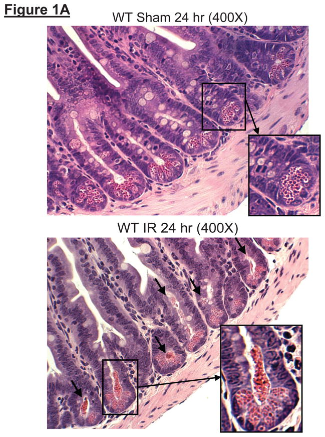
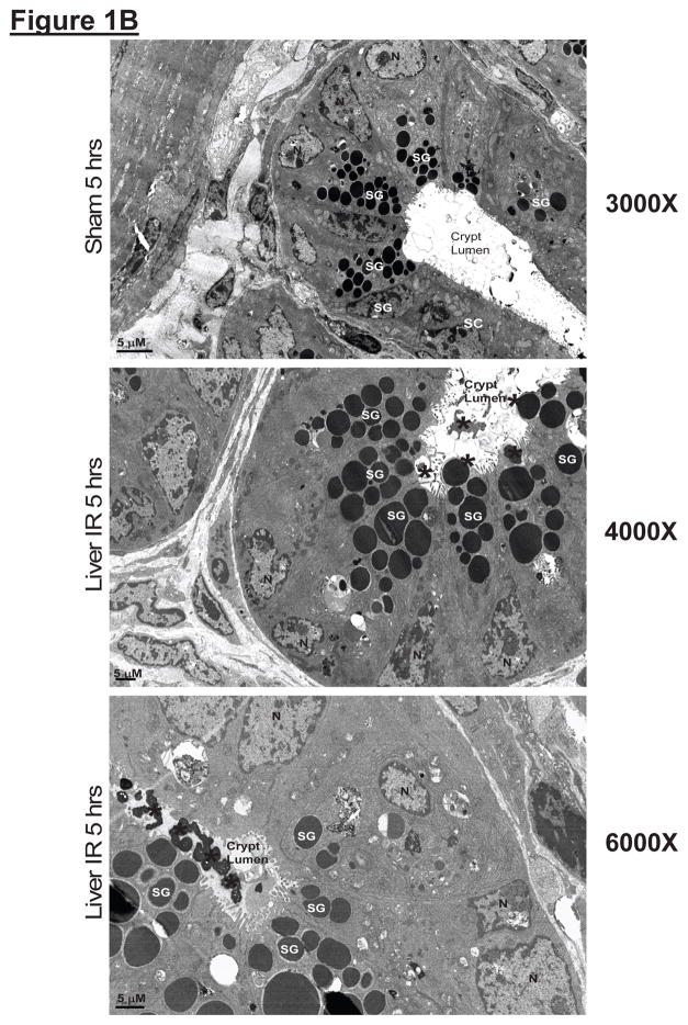
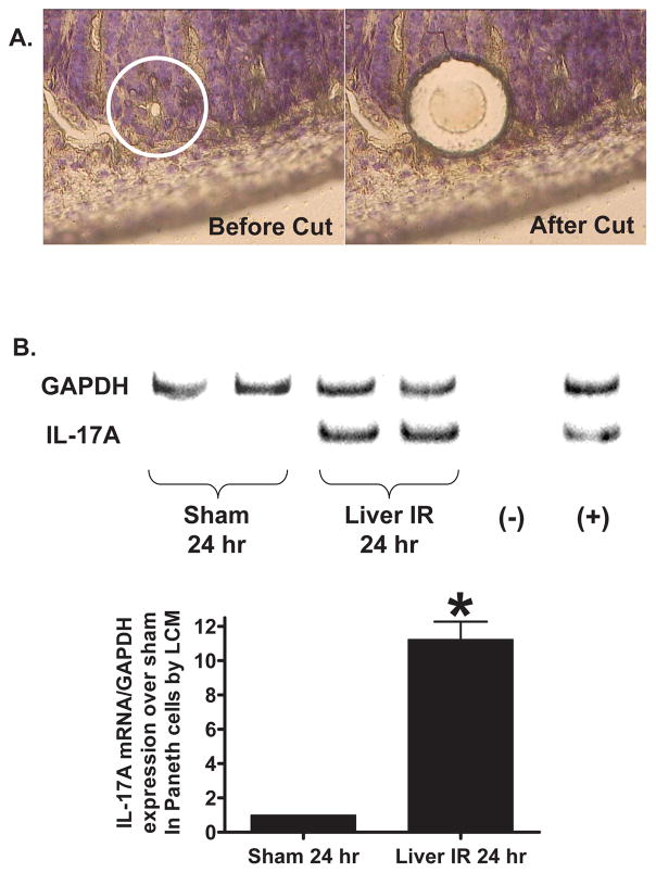
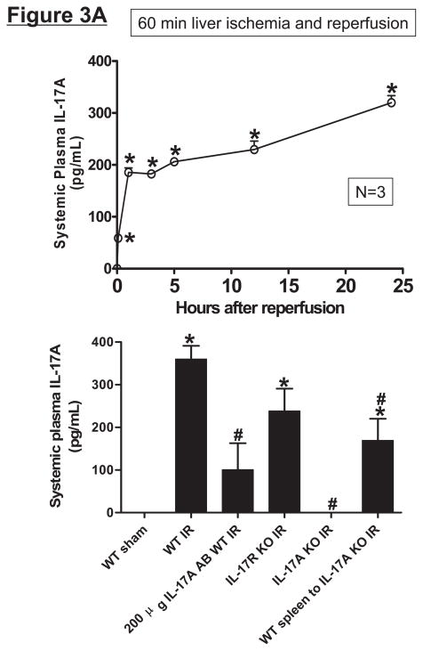
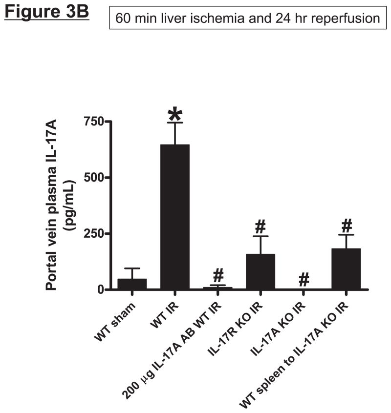
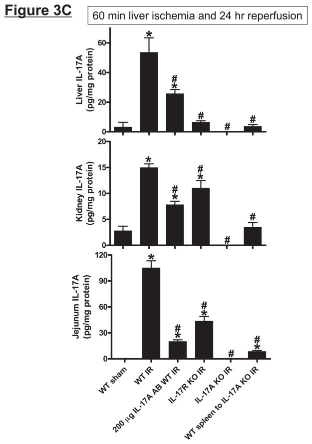
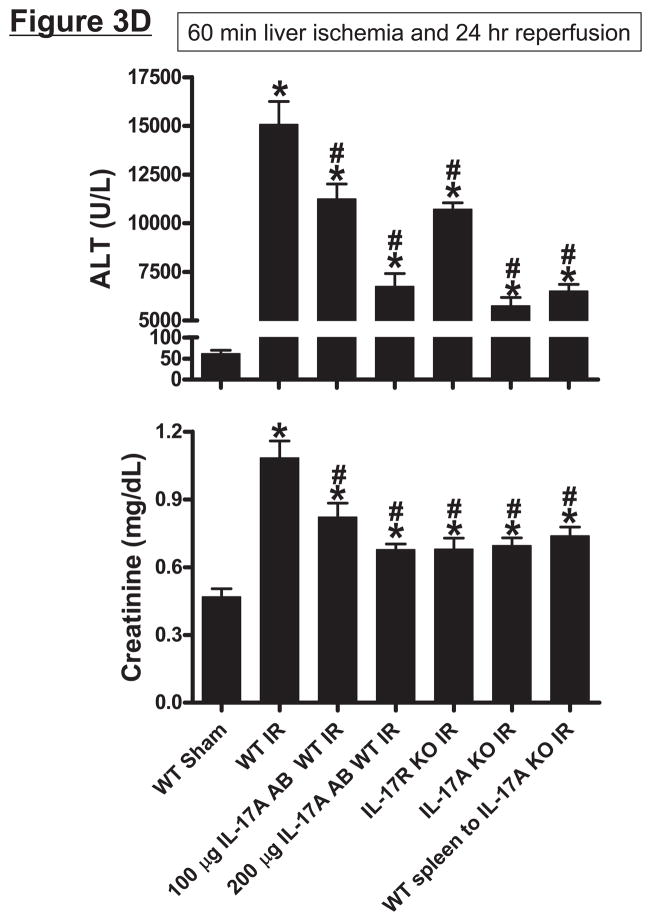
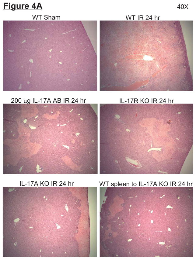
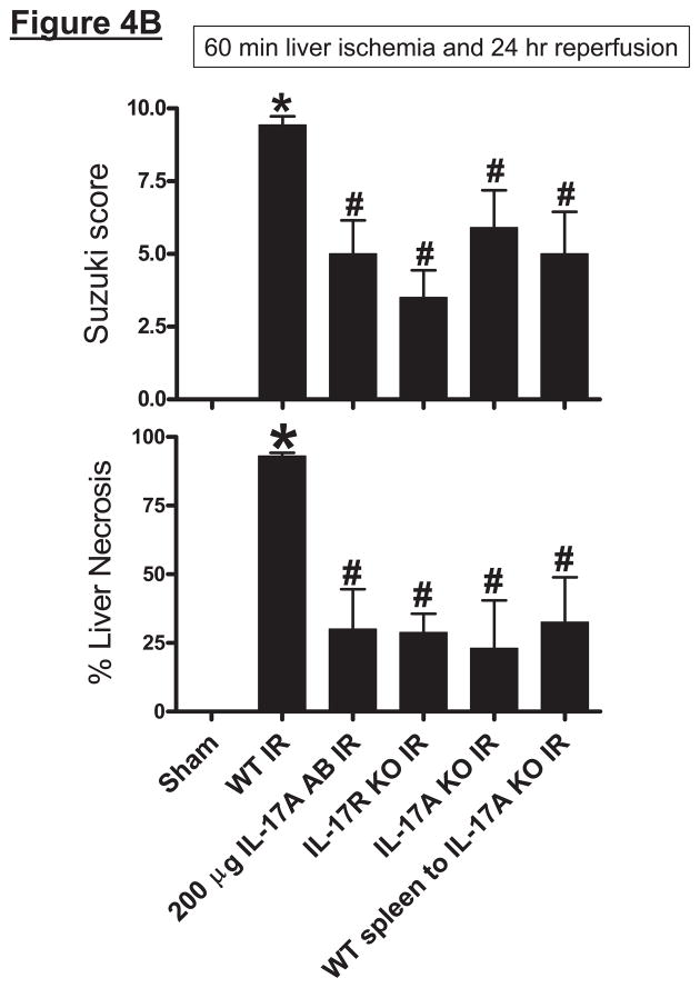
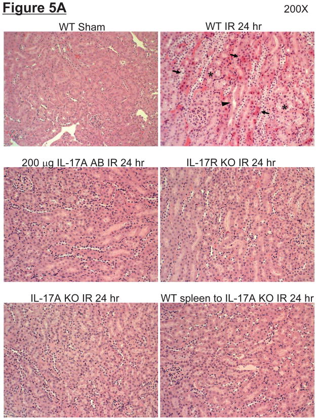
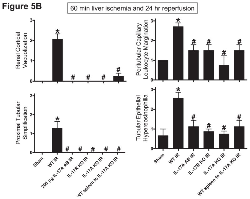
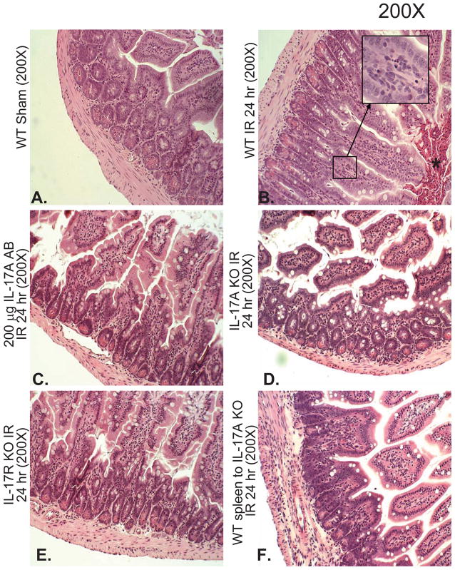
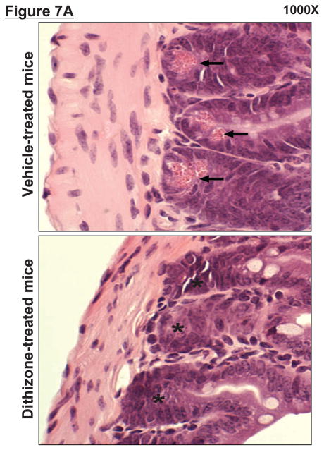
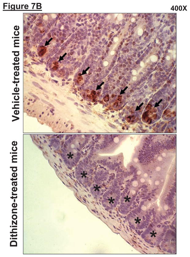
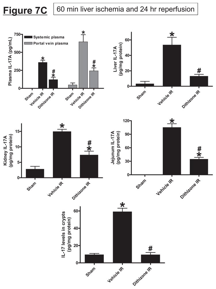
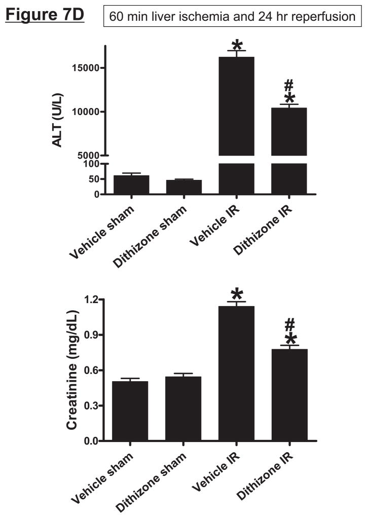
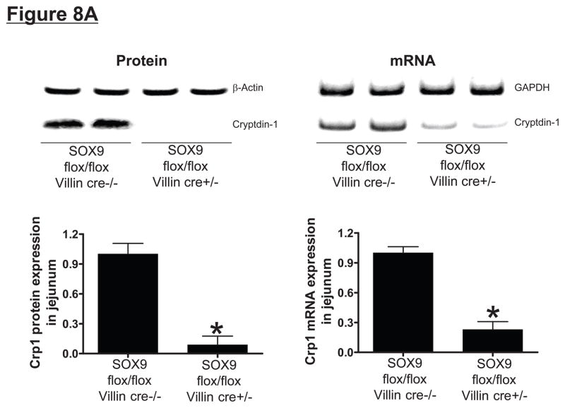
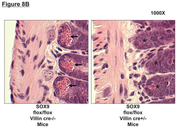
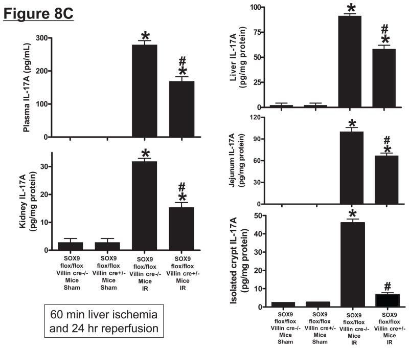
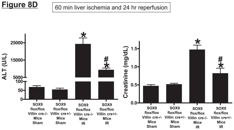
Hepatic ischemia and reperfusion (IR) injury is a major clinical problem that leads to frequent extrahepatic complications including intestinal dysfunction and acute kidney injury (AKI). In this study we aimed to determine the mechanisms of hepatic IR-induced extrahepatic organ dysfunction. Mice subjected to 60 minutes of hepatic IR not only developed severe hepatic injury but also developed significant AKI and small intestinal injury. Hepatic IR induced small intestinal Paneth cell degranulation and increased interleukin-17A (IL-17A) levels in portal vein plasma and small intestine. We also detected increased levels of IL-17A messenger RNA (mRNA) and protein in Paneth cells after hepatic IR with laser capture dissection. IL-17A-neutralizing antibody treatment or genetic deletion of either IL-17A or IL-17A receptors significantly protected against hepatic IR-induced acute liver, kidney, and intestinal injury. Leukocyte IL-17A does not contribute to organ injury, as infusion of wildtype splenocytes failed to exacerbate liver and kidney injury in IL-17A-deficient mice after hepatic IR. Depletion of Paneth cell numbers by pharmacological (with dithizone) or genetic intervention (SOX9 flox/flox Villin cre+/- mice) significantly attenuated intestinal, hepatic, and renal injury following liver IR. Finally, depletion of Paneth cell numbers significantly decreased small intestinal IL-17A release and plasma IL-17A levels after liver IR.

Conclusion: Taken together, the results show that Paneth cell-derived IL-17A plays a critical role in hepatic IR injury and extrahepatic organ dysfunction. Modulation of Paneth cell dysregulation may have therapeutic implications by reducing systemic complications arising from hepatic IR.


Conflict of interest statement

Disclosure

None of the authors had financial interests or ties to commercial companies.

Figures

Figure 1
Figure 1
A. Representative H&E staining images of small intestinal (ileum shown) Paneth cells containing dense eosinophilic granules within their apical cytoplasm (400X magnification). Hepatic IR resulted in small intestinal Paneth cell degranulation (B and C) in 5 hrs compared to sham-operated animals (A). Inserts show enlarged images of Paneth cells showing degranulation into the crypt lumen. Representative of 5 experiments. B. Representative electron micrograph images of small intestinal Paneth cell degranulation (indicated by *) 5 hrs after hepatic IR (4000X and 6000X magnification shown) compared to sham-operated mice (3000X magnification). The crypt lumen from sham-operated mice was devoid of Paneth cell granules. Representative of 3 experiments. N = nucleus of Paneth cells. SG = secretory granules of Paneth cells. SC = stem cells located above the Paneth cells.
Figure 1
Figure 1
A. Representative H&E staining images of small intestinal (ileum shown) Paneth cells containing dense eosinophilic granules within their apical cytoplasm (400X magnification). Hepatic IR resulted in small intestinal Paneth cell degranulation (B and C) in 5 hrs compared to sham-operated animals (A). Inserts show enlarged images of Paneth cells showing degranulation into the crypt lumen. Representative of 5 experiments. B. Representative electron micrograph images of small intestinal Paneth cell degranulation (indicated by *) 5 hrs after hepatic IR (4000X and 6000X magnification shown) compared to sham-operated mice (3000X magnification). The crypt lumen from sham-operated mice was devoid of Paneth cell granules. Representative of 3 experiments. N = nucleus of Paneth cells. SG = secretory granules of Paneth cells. SC = stem cells located above the Paneth cells.
Figure 2
Figure 2
A. Mouse small intestine (ileum) Paneth cells before (left) and after (right) laser capture micro-dissection (400X magnification). B. Representative RT-PCR analysis (top) and band intensity quantifications (bottom) for IL-17A mRNA extracted from Paneth cells with laser capture micro-dissection. Hepatic IR caused increased IL-17 transcripts in these cells (representative of 4 experiments). (−) negative control water blank. (+) positive control RT-PCR reaction. *P<0.05 vs. sham-operated mice. Error bars represent 1 SEM.
Figure 3
Figure 3
Systemic (A) and portal plasma IL-17A levels (B), liver, kidney and small intestine (jejunum) IL-17A levels (C) and indices of hepatic (ALT) and renal (creatinine) injury (D) from mice subjected to sham-operation (Sham, N=4) or 60 min. hepatic ischemia and reperfusion (IR, N=6). Sixty min. liver IR resulted in rapid increases in plasma IL-17A levels in mice (N=3, A). Neutralization of IL-17A (IL-17A AB, 100 or 200 μg iv, N=5 each), deficiency in IL-17A receptor (IL-17R KO, N=5) or IL-17A (IL-17A KO, N=5) reduced plasma and tissue IL-17A levels and protected against hepatic and renal injury 24 hr after 60 min. hepatic IR compared to WT mice. IL-17A deficient mice (N=5) transfused with wild type splenocytes (Wild type spleen to IL-17A KO) also had significantly lower plasma and tissue IL-17A levels and were also protected against liver and kidney injury 24 hr after 60 min. hepatic IR. *P<0.05 vs. sham-operated mice. #P<0.05 vs. WT mice subjected to hepatic IR. Error bars represent 1 SEM.
Figure 3
Figure 3
Systemic (A) and portal plasma IL-17A levels (B), liver, kidney and small intestine (jejunum) IL-17A levels (C) and indices of hepatic (ALT) and renal (creatinine) injury (D) from mice subjected to sham-operation (Sham, N=4) or 60 min. hepatic ischemia and reperfusion (IR, N=6). Sixty min. liver IR resulted in rapid increases in plasma IL-17A levels in mice (N=3, A). Neutralization of IL-17A (IL-17A AB, 100 or 200 μg iv, N=5 each), deficiency in IL-17A receptor (IL-17R KO, N=5) or IL-17A (IL-17A KO, N=5) reduced plasma and tissue IL-17A levels and protected against hepatic and renal injury 24 hr after 60 min. hepatic IR compared to WT mice. IL-17A deficient mice (N=5) transfused with wild type splenocytes (Wild type spleen to IL-17A KO) also had significantly lower plasma and tissue IL-17A levels and were also protected against liver and kidney injury 24 hr after 60 min. hepatic IR. *P<0.05 vs. sham-operated mice. #P<0.05 vs. WT mice subjected to hepatic IR. Error bars represent 1 SEM.
Figure 4
Figure 4
A. Representative photomicrographs (magnification 40X) of hematoxylin and eosin staining of the liver sections. Mice were subjected to sham-operation (sham, N=4) or to 60 min. hepatic ischemia followed by 24 hr reperfusion (IR, N=6). Necrotic hepatic tissue appears as light pink with inflammatory/vascular congestion. Photographs are representative of 4–6 independent experiments. Suzuki scores and percent liver necrosis (B) for livers from mice subjected to sham or hepatic IR. Neutralization of IL-17A (IL-17A AB, 200 μg iv), deficiency in IL-17A receptor (IL-17R KO) or IL-17A (IL-17A KO) significantly reduced liver necrosis 24 hr after 60 min. hepatic IR. IL-17A deficient mice transfused with wild type splenocytes are also protected against liver injury 24 hr after 60 min. hepatic IR. *P<0.05 vs. sham-operated mice. #P<0.05 vs. mice subjected to hepatic IR. Error bars represent 1 SEM.
Figure 4
Figure 4
A. Representative photomicrographs (magnification 40X) of hematoxylin and eosin staining of the liver sections. Mice were subjected to sham-operation (sham, N=4) or to 60 min. hepatic ischemia followed by 24 hr reperfusion (IR, N=6). Necrotic hepatic tissue appears as light pink with inflammatory/vascular congestion. Photographs are representative of 4–6 independent experiments. Suzuki scores and percent liver necrosis (B) for livers from mice subjected to sham or hepatic IR. Neutralization of IL-17A (IL-17A AB, 200 μg iv), deficiency in IL-17A receptor (IL-17R KO) or IL-17A (IL-17A KO) significantly reduced liver necrosis 24 hr after 60 min. hepatic IR. IL-17A deficient mice transfused with wild type splenocytes are also protected against liver injury 24 hr after 60 min. hepatic IR. *P<0.05 vs. sham-operated mice. #P<0.05 vs. mice subjected to hepatic IR. Error bars represent 1 SEM.
Figure 5
Figure 5
A. Representative photomicrographs (magnification 200X) of hematoxylin and eosin staining of the kidney sections. Mice were subjected to sham-operation (sham, N=4) or to 60 min. hepatic ischemia followed by 24 hr reperfusion (IR, N=6). Sixty min. hepatic IR caused multifocal acute tubular injury including S3 segment proximal tubule necrosis (arrows), cortical tubular simplification (arrow head), cytoplasmic vacuolization (*). Photographs are representative of 4–6 independent experiments. B. Summary of renal injury scores (scale 0–3) for renal cortical vacuolization, peritubular leukocyte margination, proximal tubule simplification and renal tubular hypereosinophilia for kidney sections from mice subjected to sham or hepatic IR. Neutralization of IL-17A (IL-17A AB, 200 μg iv), deficiency in IL-17A receptor (IL-17R KO) or IL-17A (IL-17A KO) significantly reduces the renal injury 24 hr after 60 min. hepatic IR. IL-17A deficient mice transfused with wild type splenocytes are also protected against the renal injury 24 hr after 60 min. hepatic IR. *P<0.05 vs. sham-operated mice. #P<0.05 vs. mice subjected to hepatic IR. Error bars represent 1 SEM.
Figure 5
Figure 5
A. Representative photomicrographs (magnification 200X) of hematoxylin and eosin staining of the kidney sections. Mice were subjected to sham-operation (sham, N=4) or to 60 min. hepatic ischemia followed by 24 hr reperfusion (IR, N=6). Sixty min. hepatic IR caused multifocal acute tubular injury including S3 segment proximal tubule necrosis (arrows), cortical tubular simplification (arrow head), cytoplasmic vacuolization (*). Photographs are representative of 4–6 independent experiments. B. Summary of renal injury scores (scale 0–3) for renal cortical vacuolization, peritubular leukocyte margination, proximal tubule simplification and renal tubular hypereosinophilia for kidney sections from mice subjected to sham or hepatic IR. Neutralization of IL-17A (IL-17A AB, 200 μg iv), deficiency in IL-17A receptor (IL-17R KO) or IL-17A (IL-17A KO) significantly reduces the renal injury 24 hr after 60 min. hepatic IR. IL-17A deficient mice transfused with wild type splenocytes are also protected against the renal injury 24 hr after 60 min. hepatic IR. *P<0.05 vs. sham-operated mice. #P<0.05 vs. mice subjected to hepatic IR. Error bars represent 1 SEM.
Figure 6
Figure 6
A. Representative photomicrographs of small intestine from 4–6 experiments (hematoxylin and eosin staining, magnification 200X) of mice subjected to sham-operation (sham, N=4) or to 60 min. hepatic ischemia followed by 24 hr reperfusion (IR, N=6). Sham operated animals show normal-appearing intestine histology (A). In contrast, the small intestine sections from mice subjected to hepatic IR show villous endothelial cell apoptosis (magnified insert), severe epithelial cell necrosis of villous lining cells and the development of a necrotic epithelial pannus (*) over the mucosal surface compared to sham-operated animals (B). Neutralization of IL-17A (IL-17A AB, 200 μg iv, C), deficiency in IL-17A (IL-17S KO, D) or IL-17A receptor (IL-17R KO, E) significantly reduces the intestine injury 24 hr after 60 min. hepatic IR. IL-17A deficient mice transfused with wild type splenocytes are also protected against the intestine injury 24 hr after 60 min. hepatic IR (F).
Figure 7
Figure 7
A. Dithizone treatment depletes small intestinal Paneth cell granules (*). Representative (of 4 experiments) hematoxylin and eosin staining images of ileum from mice treated with vehicle (Li2CO3) or with dithizone 6 hr prior. Note near complete depletion of Paneth cell granules (arrows, magnification 1000X) after dithizone treatment (*). B. Representative (of 5 independent experiments, magnification 400X) images of lysozyme immunostaining in small intestine (ileum). Note that lysozyme stain was heavy in Paneth cells (arrows) of small intestinal crypts of mice treated with vehicle (LiCO3). Paneth cell depletion with dithizone treatment eliminated lysozyme staining in Paneth cells (*). C. Dithizone treatment reduces plasma IL-17A levels in mice subjected to 60 min. hepatic IR (Dithizone IR, N=4). Dithizone treatment also reduced IL-17A protein upregulation in liver, kidney, small intestine and freshly isolated small intestinal crypts (N=4) 24 hr after 60 min. hepatic IR. D. Paneth cell granule depletion with dithizone treatment protects against hepatic (ALT) and renal (creatinine) injury after liver IR. Mice were subjected to sham-operation (Sham, N=4) or hepatic IR (N=6), and plasma was collected 24 hrs after reperfusion. *P<0.05 vs. sham-operated mice. #P<0.05 vs. vehicle treated mice subjected to hepatic IR. Error bars represent 1 SEM.
Figure 7
Figure 7
A. Dithizone treatment depletes small intestinal Paneth cell granules (*). Representative (of 4 experiments) hematoxylin and eosin staining images of ileum from mice treated with vehicle (Li2CO3) or with dithizone 6 hr prior. Note near complete depletion of Paneth cell granules (arrows, magnification 1000X) after dithizone treatment (*). B. Representative (of 5 independent experiments, magnification 400X) images of lysozyme immunostaining in small intestine (ileum). Note that lysozyme stain was heavy in Paneth cells (arrows) of small intestinal crypts of mice treated with vehicle (LiCO3). Paneth cell depletion with dithizone treatment eliminated lysozyme staining in Paneth cells (*). C. Dithizone treatment reduces plasma IL-17A levels in mice subjected to 60 min. hepatic IR (Dithizone IR, N=4). Dithizone treatment also reduced IL-17A protein upregulation in liver, kidney, small intestine and freshly isolated small intestinal crypts (N=4) 24 hr after 60 min. hepatic IR. D. Paneth cell granule depletion with dithizone treatment protects against hepatic (ALT) and renal (creatinine) injury after liver IR. Mice were subjected to sham-operation (Sham, N=4) or hepatic IR (N=6), and plasma was collected 24 hrs after reperfusion. *P<0.05 vs. sham-operated mice. #P<0.05 vs. vehicle treated mice subjected to hepatic IR. Error bars represent 1 SEM.
Figure 8
Figure 8
SOX9 flox/flox Villin Cre+/− (selective SOX9 deletion in intestinal epithelia) mice are deficient in Paneth cell marker (cryptdin-1 protein and mRNA, A) and in Paneth cells (B) compared to wild type (SOX9 flox/flox Villin Cre−/−) mice. Figure 7B shows near complete deficiency of Paneth cells (indicated by arrows in SOX WT mice, magnification 1000X) in SOX9 flox/flox Villin Cre+/− mice (*). C. Mice were subjected to sham-operation (Sham, N=4) or 60 min. hepatic IR (IR, N=4). Paneth cell deficiency in SOX9 flox/flox Villin Cre+/− mice reduces plasma, liver, kidney, small intestine (jejunum shown) and freshly isolated small intestinal crypt IL-17A levels in mice subjected to 60 min. hepatic IR (N=4). D. Paneth cell deficiency in SOX9 flox/flox Villin Cre+/− mice protects against hepatic (ALT) and renal (creatinine) injury compared with SOX9 flox/flox Villin Cre−/− mice subjected 60 min. hepatic IR. *P<0.05 vs. sham-operated WT mice. #P<0.05 vs. WT mice subjected to hepatic IR. Error bars represent 1 SEM.
Figure 8
Figure 8
SOX9 flox/flox Villin Cre+/− (selective SOX9 deletion in intestinal epithelia) mice are deficient in Paneth cell marker (cryptdin-1 protein and mRNA, A) and in Paneth cells (B) compared to wild type (SOX9 flox/flox Villin Cre−/−) mice. Figure 7B shows near complete deficiency of Paneth cells (indicated by arrows in SOX WT mice, magnification 1000X) in SOX9 flox/flox Villin Cre+/− mice (*). C. Mice were subjected to sham-operation (Sham, N=4) or 60 min. hepatic IR (IR, N=4). Paneth cell deficiency in SOX9 flox/flox Villin Cre+/− mice reduces plasma, liver, kidney, small intestine (jejunum shown) and freshly isolated small intestinal crypt IL-17A levels in mice subjected to 60 min. hepatic IR (N=4). D. Paneth cell deficiency in SOX9 flox/flox Villin Cre+/− mice protects against hepatic (ALT) and renal (creatinine) injury compared with SOX9 flox/flox Villin Cre−/− mice subjected 60 min. hepatic IR. *P<0.05 vs. sham-operated WT mice. #P<0.05 vs. WT mice subjected to hepatic IR. Error bars represent 1 SEM.

References

    1. Kupiec-Weglinski JW, Busuttil RW. Ischemia and reperfusion injury in liver transplantation. Transplant Proc. 2005 May;37(4):1653–1656. - PubMed
    1. Davis CL, Gonwa TA, Wilkinson AH. Pathophysiology of renal disease associated with liver disorders: implications for liver transplantation. Part I. Liver Transpl. 2002 Feb;8(2):91–109. - PubMed
    1. Cua DJ, Tato CM. Innate IL-17-producing cells: the sentinels of the immune system. Nat Rev Immunol. 2010 Jul;10(7):479–489. - PubMed
    1. Takahashi N, Vanlaere I, de RR, Cauwels A, Joosten LA, Lubberts E, et al. IL-17 produced by Paneth cells drives TNF-induced shock. J Exp Med. 2008 Aug 4;205(8):1755–1761. - PMC - PubMed
    1. Chang SH, Dong C. IL-17F: regulation, signaling and function in inflammation. Cytokine. 2009 Apr;46(1):7–11. - PMC - PubMed

Publication types