. 2011 Aug;66(2):428-36.
doi: 10.1002/mrm.22814. Epub 2011 Feb 28.

Interleaved variable density sampling with a constrained parallel imaging reconstruction for dynamic contrast-enhanced MR angiography

Affiliations

Interleaved variable density sampling with a constrained parallel imaging reconstruction for dynamic contrast-enhanced MR angiography

Kang Wang et al. Magn Reson Med. 2011 Aug.

Abstract

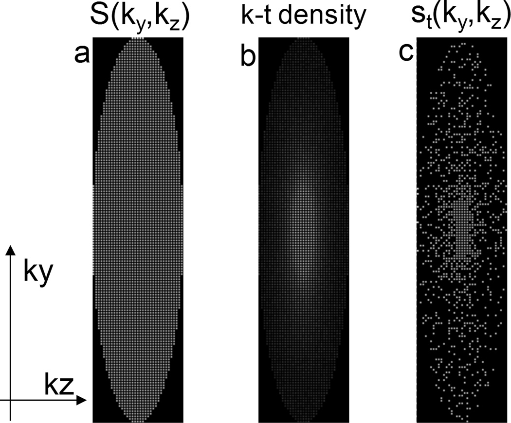
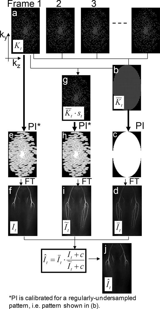
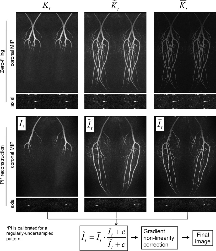
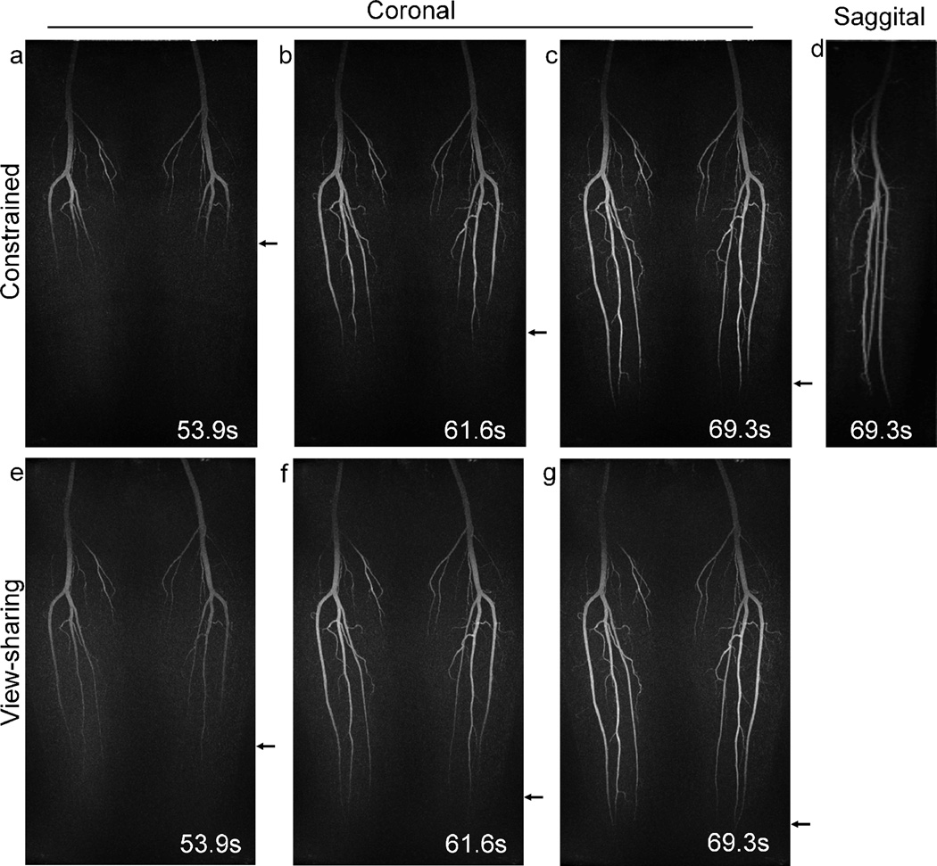
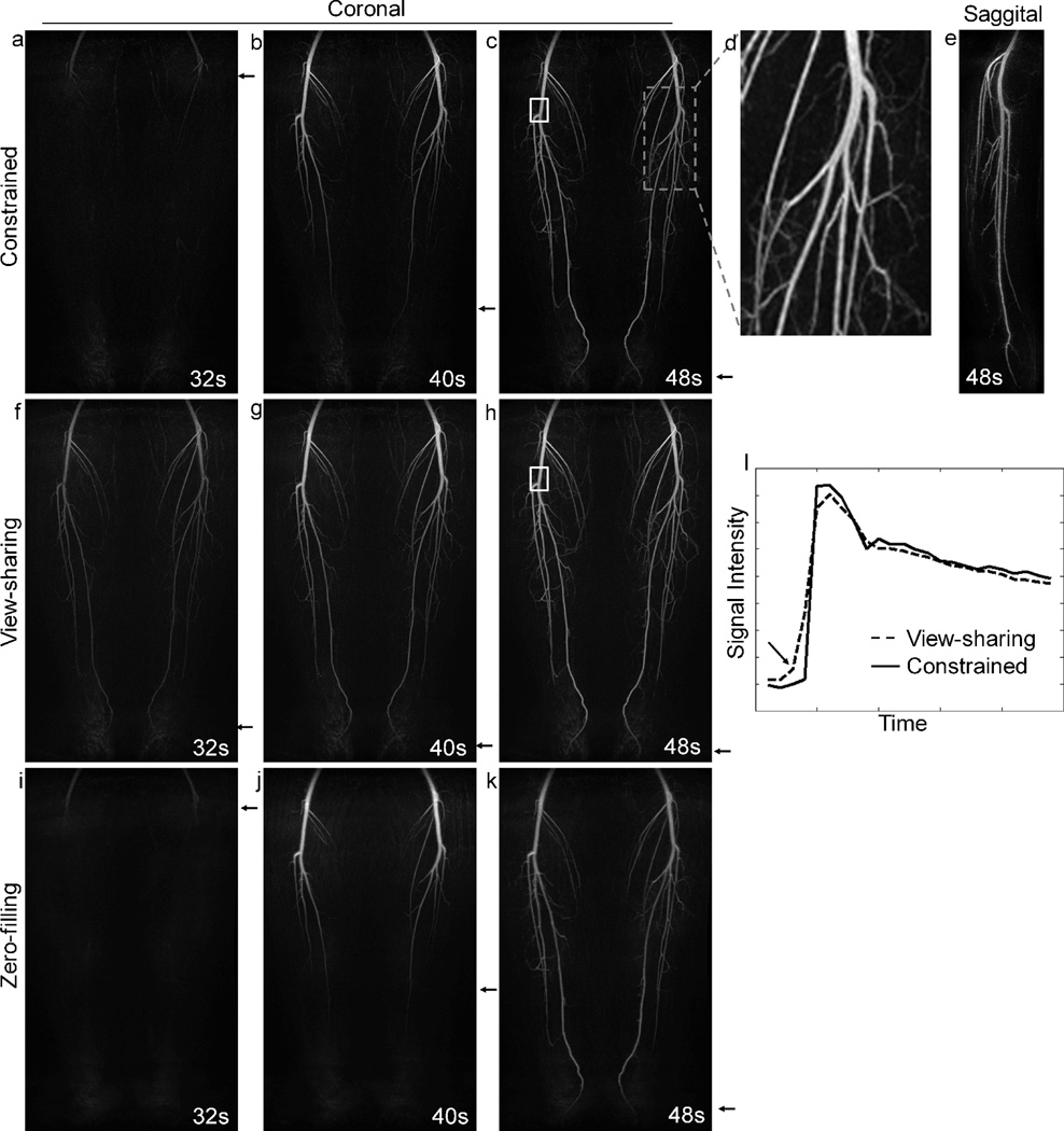
For MR applications such as contrast-enhanced MR angiography, it is desirable to achieve simultaneously high spatial and temporal resolution. The current clinical standard uses view-sharing methods combined with parallel imaging; however, this approach still provides limited spatial and temporal resolution. To improve on the clinical standard, we present an interleaved variable density (IVD) sampling method that pseudorandomly undersamples each individual frame of a 3D Cartesian ky-kz plane combined with parallel imaging acceleration. From this dataset, time-resolved images are reconstructed with a method that combines parallel imaging with a multiplicative constraint. Total acceleration factors on the order of 20 are achieved for contrast-enhanced MR angiography of the lower extremities, and improvements in temporal fidelity of the depiction of the contrast bolus passage are demonstrated relative to the clinical standard.


Figures

Figure 1
Figure 1
(a) All view locations that are sampled during the course of an acquisition, with parallel imaging acceleration factor of 4. (b) A given k-t sampling density (1/kr) over N time frames. (c) An example of the IVD sampling pattern for an individual frame with an additional acceleration of 4 (total acceleration of 16).
Figure 2
Figure 2
Diagram for the Cartesian constrained parallel imaging reconstruction method. Step 1:(a,b) Data from a subset of all the time frames are combined, using a sliding window average to create a composite data set that has uniform undersampling on the 2 × 2 acceleration grid, t. (c) parallel imaging is used to synthesize the missing data points. (d) An FFT of these data produces the temporally-blurred, high spatial resolution, high SNR image, Īt. Step 2:(e) For data in each undersampled time frame Kt, parallel imaging is used to synthesize points off the 2 × 2 acceleration grid. (f) An FFT of these data produce the high temporal resolution, low spatial resolution, high SNR image It. Step 3:(g) The t are subsampled using the IVD pattern for that time frame t. (h) parallel imaging is used to synthesize points off the 2 × 2 acceleration grid. (i) An FFT of these data produce the image Ĩt. Step 4:(j) The final image Ît is calculated using Eq. [2].
Figure 3
Figure 3
Intermediate images in the proposed reconstruction method. Images in the first row are reconstructed using zero-filling and exhibit coherent aliasing artifacts due to the regular undersampling by parallel imaging. Images in the second row are reconstructed using parallel imaging calibrated for a regularly-undersampled pattern. These images do not have coherent aliasing (parallel imaging) artifacts, although images It and Ĩt exhibit expected spatial blurring and incoherent artifacts due to the IVD undersampling. However, these artifacts can be suppressed in the constrained reconstruction in the next step (bottom equation). Note that although images shown here are coil-combined (for demonstration purpose), the multiplicative constrained reconstruction is applied in a coil-by-coil basis (see Discussion). Also notice that the compensation for non-linear gradient effects is not applied to the intermediate images shown here; instead it is only applied to the image Ît after the constrained reconstruction and coil combination.
Figure 4
Figure 4
Results obtained from volunteer 1. The constrained reconstruction method implemented in conjunction with parallel imaging shows better depiction of the progressive filling of the popliteal and tibial arteries than the view-sharing method, as indicated by the arrows. A sagittal reformat (d) of the constrained reconstruction image confirms that no aliasing is present.
Figure 5
Figure 5
Results obtained from volunteer 2. (a–c) Coronal MIP images from the constrained reconstruction showing good depiction of the arteries filling with contrast material. (d) An enlarged image of the dashed box with small arteries well visualized. (e): sagittal reformat. (f–h) Same data set reconstructed using view-sharing method and parallel imaging. (i–k) Same data set reconstructed using zero-filled parallel imaging calibrated for a regularly-undersampled pattern. Note that no apparent coherent aliasing are present in these images. (l) Waveforms obtained from view-sharing and constrained reconstruction, with the former method causing temporal blurring indicated by the arrow.

Similar articles

Cited by

References

    1. Zhang H, Maki JH, Prince MR. 3D contrast-enhanced MR angiography. J Magn Reson Imaging. 2007;25(1):13–25. - PubMed
    1. Haider CR, Hu HH, Campeau NG, Huston J, 3rd, Riederer SJ. 3D high temporal and spatial resolution contrast-enhanced MR angiography of the whole brain. Magn Reson Med. 2008;60(3):749–760. - PMC - PubMed
    1. Haider CR, Glockner JF, Vrtiska TJ, Macedo TA, Borisch EA, Riederer SJ. A Comparison of Time-Resolved 3D CE-MRA with Peripheral Run-off CTA in the Calves. East Lansing, MI: MR Angio Club; 2009.
    1. Du J. Contrast-enhanced MR angiography using time resolved interleaved projection sampling with three-dimensional cartesian phase and slice encoding (TRIPPS) Magn Reson Med. 2009;61(4):918–924. - PubMed
    1. Lustig M, Donoho D, Pauly JM. Sparse MRI: The application of compressed sensing for rapid MR imaging. Magn Reson Med. 2007;58(6):1182–1195. - PubMed

Publication types

MeSH terms