. 2011 Jan;38(1):228-37.
doi: 10.1118/1.3525835.

Segmentation and quantification of materials with energy discriminating computed tomography: a phantom study

Affiliations

Segmentation and quantification of materials with energy discriminating computed tomography: a phantom study

Huy Q Le et al. Med Phys. 2011 Jan.

Abstract

Purpose: To experimentally investigate whether a computed tomography (CT) system based on CdZnTe (CZT) detectors in conjunction with a least-squares parameter estimation technique can be used to decompose four different materials.

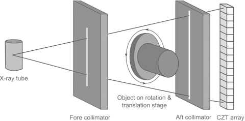
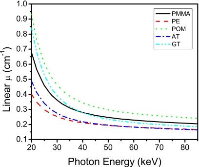
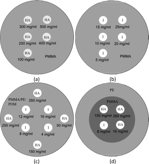
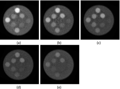
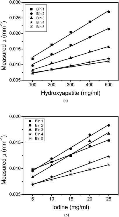
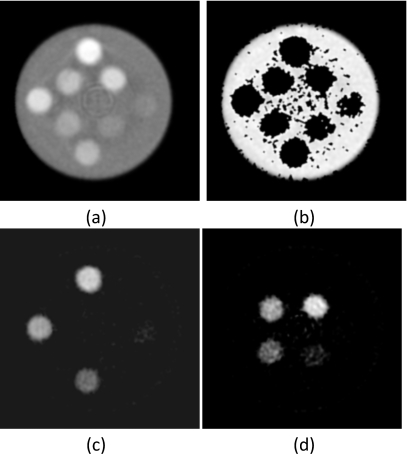
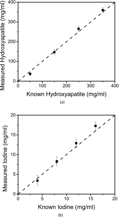
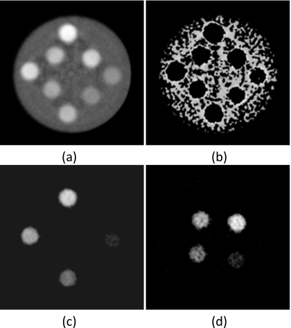
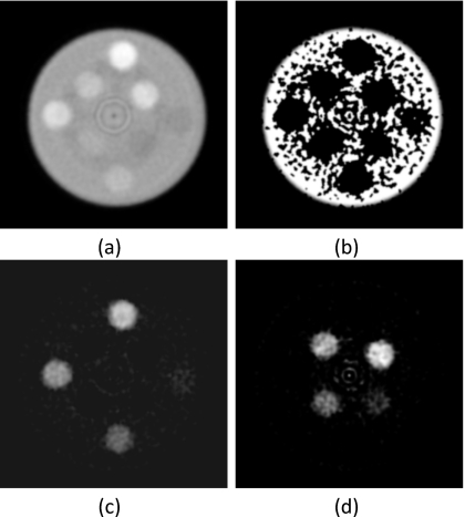
Methods: The material decomposition process was divided into a segmentation task and a quantification task. A least-squares minimization algorithm was used to decompose materials with five measurements of the energy dependent linear attenuation coefficients. A small field-of-view energy discriminating CT system was built. The CT system consisted of an x-ray tube, a rotational stage, and an array of CZT detectors. The CZT array was composed of 64 pixels, each of which is 0.8 x 0.8 x 3 mm. Images were acquired at 80 kVp in fluoroscopic mode at 50 ms per frame. The detector resolved the x-ray spectrum into energy bins of 22-32, 33-39, 40-46, 47-56, and 57-80 keV. Four phantoms were constructed from polymethylmethacrylate (PMMA), polyethylene, polyoxymethylene, hydroxyapatite, and iodine. Three phantoms were composed of three materials with embedded hydroxyapatite (50, 150, 250, and 350 mg/ml) and iodine (4, 8, 12, and 16 mg/ml) contrast elements. One phantom was composed of four materials with embedded hydroxyapatite (150 and 350 mg/ml) and iodine (8 and 16 mg/ml). Calibrations consisted of PMMA phantoms with either hydroxyapatite (100, 200, 300, 400, and 500 mg/ml) or iodine (5, 15, 25, 35, and 45 mg/ml) embedded. Filtered backprojection and a ramp filter were used to reconstruct images from each energy bin. Material segmentation and quantification were performed and compared between different phantoms.

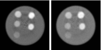
Results: All phantoms were decomposed accurately, but some voxels in the base material regions were incorrectly identified. Average quantification errors of hydroxyapatite/iodine were 9.26/7.13%, 7.73/5.58%, and 12.93/8.23% for the three-material PMMA, polyethylene, and polyoxymethylene phantoms, respectively. The average errors for the four-material phantom were 15.62% and 2.76% for hydroxyapatite and iodine, respectively.

Conclusions: The calibrated least-squares minimization technique of decomposition performed well in breast imaging tasks with an energy resolving detector. This method can provide material basis images containing concentrations of the relevant materials that can potentially be valuable in the diagnostic process.


Figures

Figure 1
Figure 1
Schematic of the CZT system.
Figure 2
Figure 2
Linear attenuation coefficients of the plastics used in the base material of the phantoms. Adipose (AT) and glandular (GT) tissues are also shown for comparison. PMMA=polymethylmethacrylate, PE=polyethylene, and POM=polyoxymethylene.
Figure 3
Figure 3
Diagrams of the calibration phantoms with (a) hydroxyapatite and (b) iodine contrast elements and measurement phantoms of (c) three and (d) four materials. PMMA=polymethylmethacrylate, PE=polyethylene, POM=polyoxymethylene, HA=hydroxyapatite, and I=iodine.
Figure 4
Figure 4
Photon counting CT images of hydroxyapatite calibration (left) and iodine calibration (right) phantoms (L∕W:0.010∕0.02 mm−1).
Figure 5
Figure 5
CT slices of the polymethylmethacrylate three-material phantom for energy bins (a) 1, (b) 2, (c) 3, (d) 4, and (e) 5 (L∕W:0.01∕0.02 mm−1).
Figure 6
Figure 6
Calibration curves for (a) HA and (b) iodine. The slope of the each line indicates the effective mass attenuation coefficients for the corresponding energy bin.
Figure 7
Figure 7
Material separation was applied to the polymethylmethacrylate three-material phantom. (a) Photon counting image (L∕W:0.01∕0.01 mm−1) and decomposed images of (b) PMMA (L∕W: 600∕1000 mg∕ml), (c) hydroxyapatite (L∕W: 200∕600 mg∕ml), and (d) iodine (L∕W: 10∕20 mg∕ml) are shown.
Figure 8
Figure 8
Relationships between the measured and known concentrations of (a) hydroxyapatite and (b) iodine. The identity line is shown is dashed.
Figure 9
Figure 9
Material separation was performed on the polyethylene three-material phantom. (a) Photon counting image (L∕W:0.01∕0.01 mm−1) and decomposed images of (b) PMMA (L∕W: 600∕1000 mg∕ml), (c) hydroxyapatite (L∕W: 200∕600 mg∕ml), and (d) iodine (L∕W: 10∕20 mg∕ml) are shown. Base material was calibrated with PMMA.
Figure 10
Figure 10
Material separation was performed on the polyoxymethylene three-material phantom. (a) Photon counting image (L∕W:0.01∕0.01 mm−1) and decomposed images of (b) PMMA (L∕W: 600∕1000 mg∕ml), (c) hydroxyapatite (L∕W: 200∕600 mg∕ml), and (d) iodine (L∕W: 10∕20 mg∕ml) are shown. Base material was calibrated with PMMA.
Figure 11
Figure 11
Material separation was performed on the four-material phantom. (a) Photon counting image (L∕W:0.01∕0.01 mm−1) and decomposed images of (b) PE (L∕W: 600∕1000 mg∕ml), (c) PMMA (L∕W: 600∕1000 mg∕ml), (d) hydroxyapatite (L∕W: 200∕600 mg∕ml), (e) and iodine (L∕W: 10∕20 mg∕ml) are shown.

References

    1. Alvarez R. E. and Macovski A., “Energy-selective reconstructions in x-ray computerized tomography,” Phys. Med. Biol. PHMBA7 21, 733–744 (1976).10.1088/0031-9155/21/5/002 - DOI - PubMed
    1. Liu X., Yu L., Primak A. N., and McCollough C. H., “Quantitative imaging of element composition and mass fraction using dual-energy CT: Three-material decomposition,” Med. Phys. MPHYA6 36, 1602–1609 (2009).10.1118/1.3097632 - DOI - PMC - PubMed
    1. Lehmann L. A., Alvarez R. E., Macovski A., Brody W. R., Pelc N. J., Riederer S. J., and Hall A. L., “Generalized image combinations in dual KVP digital radiography,” Med. Phys. MPHYA6 8, 659–667 (1981).10.1118/1.595025 - DOI - PubMed
    1. Kalender W. A., Perman W. H., Vetter J. R., and Klotz E., “Evaluation of a prototype dual-energy computed tomographic apparatus. 1. Phantom studies,” Med. Phys. MPHYA6 13, 334–339 (1986).10.1118/1.595958 - DOI - PubMed
    1. Heismann B. J., Leppert J., and Stierstorfer K., “Density and atomic number measurements with spectral x-ray attenuation method,” J. Appl. Phys. JAPIAU 94, 2073–2079 (2003).10.1063/1.1586963 - DOI

Publication types