Patient-specific radiation dose and cancer risk estimation in CT: part II. Application to patients
- PMID: 21361209
- PMCID: PMC3021563
- DOI: 10.1118/1.3515864
Patient-specific radiation dose and cancer risk estimation in CT: part II. Application to patients
Abstract
Purpose: Current methods for estimating and reporting radiation dose from CT examinations are largely patient-generic; the body size and hence dose variation from patient to patient is not reflected. Furthermore, the current protocol designs rely on dose as a surrogate for the risk of cancer incidence, neglecting the strong dependence of risk on age and gender. The purpose of this study was to develop a method for estimating patient-specific radiation dose and cancer risk from CT examinations.
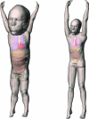
Methods: The study included two patients (a 5-week-old female patient and a 12-year-old male patient), who underwent 64-slice CT examinations (LightSpeed VCT, GE Healthcare) of the chest, abdomen, and pelvis at our institution in 2006. For each patient, a nonuniform rational B-spine (NURBS) based full-body computer model was created based on the patient's clinical CT data. Large organs and structures inside the image volume were individually segmented and modeled. Other organs were created by transforming an existing adult male or female full-body computer model (developed from visible human data) to match the framework defined by the segmented organs, referencing the organ volume and anthropometry data in ICRP Publication 89. A Monte Carlo program previously developed and validated for dose simulation on the LightSpeed VCT scanner was used to estimate patient-specific organ dose, from which effective dose and risks of cancer incidence were derived. Patient-specific organ dose and effective dose were compared with patient-generic CT dose quantities in current clinical use: the volume-weighted CT dose index (CTDIvol) and the effective dose derived from the dose-length product (DLP).
Results: The effective dose for the CT examination of the newborn patient (5.7 mSv) was higher but comparable to that for the CT examination of the teenager patient (4.9 mSv) due to the size-based clinical CT protocols at our institution, which employ lower scan techniques for smaller patients. However, the overall risk of cancer incidence attributable to the CT examination was much higher for the newborn (2.4 in 1000) than for the teenager (0.7 in 1000). For the two pediatric-aged patients in our study, CTDIvol underestimated dose to large organs in the scan coverage by 30%-48%. The effective dose derived from DLP using published conversion coefficients differed from that calculated using patient-specific organ dose values by -57% to 13%, when the tissue weighting factors of ICRP 60 were used, and by -63% to 28%, when the tissue weighting factors of ICRP 103 were used.
Conclusions: It is possible to estimate patient-specific radiation dose and cancer risk from CT examinations by combining a validated Monte Carlo program with patient-specific anatomical models that are derived from the patients' clinical CT data and supplemented by transformed models of reference adults. With the construction of a large library of patient-specific computer models encompassing patients of all ages and weight percentiles, dose and risk can be estimated for any patient prior to or after a CT examination. Such information may aid in decisions for image utilization and can further guide the design and optimization of CT technologies and scan protocols.
Figures


References
-
- Hurwitz L. M., Yoshizumi T. T., Goodman P. C., Nelson R. C., Toncheva G., Nguyen G. B., Lowry C., and Anderson-Evans C., “Radiation dose savings for adult pulmonary embolus 64-MDCT using bismuth breast shields, lower peak kilovoltage, and automatic tube current modulation,” AJR, Am. J. Roentgenol. AAJRDX 192, 244–253 (2009).10.2214/AJR.08.1066 - DOI - PubMed
-
- Cristy M. and Eckerman K. F., “Specific absorbed fractions of energy at various ages from internal photon sources,” Oak Ridge National Laboratory Report No. ORNL/TM-8381.

