Review
. 2011 Mar-Apr;19(2):134-48.
doi: 10.1111/j.1524-475X.2011.00673.x.

Dynamic reciprocity in the wound microenvironment

Affiliations
Review

Dynamic reciprocity in the wound microenvironment

Gregory S Schultz et al. Wound Repair Regen. 2011 Mar-Apr.

Abstract

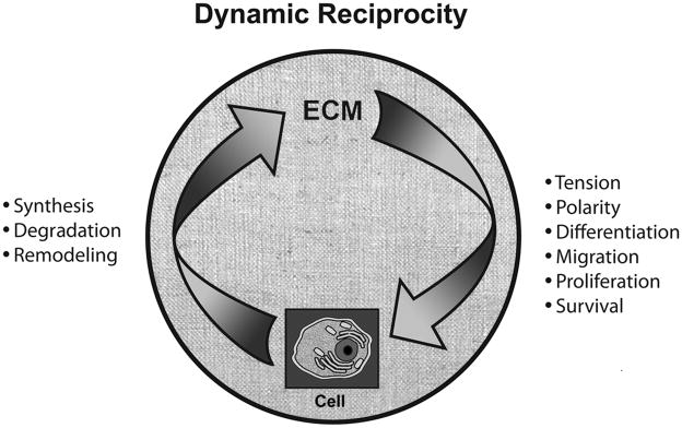
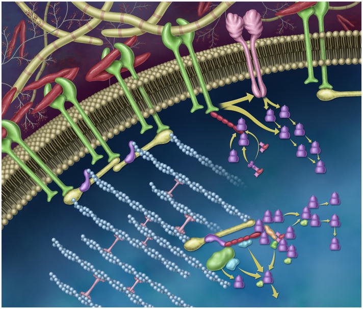
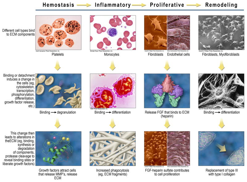
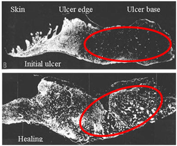
Here, we define dynamic reciprocity (DR) as an ongoing, bidirectional interaction among cells and their surrounding microenvironment. In this review, we posit that DR is especially meaningful during wound healing as the DR-driven biochemical, biophysical, and cellular responses to injury play pivotal roles in regulating tissue regenerative responses. Such cell-extracellular matrix interactions not only guide and regulate cellular morphology, but also cellular differentiation, migration, proliferation, and survival during tissue development, including, e.g., embryogenesis, angiogenesis, as well as during pathologic processes including cancer, diabetes, hypertension, and chronic wound healing. Herein, we examine DR within the wound microenvironment while considering specific examples across acute and chronic wound healing. This review also considers how a number of hypotheses that attempt to explain chronic wound pathophysiology may be understood within the DR framework. The implications of applying the principles of DR to optimize wound care practice and future development of innovative wound healing therapeutics are also briefly considered.


Figures

Figure 1
Figure 1
DR between cells and ECM. Cells synthesize ECM components, and also degrade and remodel ECM, the latter events occurring through the production and regulation of matrix metalloproteases (MMPs) and other enzymes. The ECM regulates cellular tension and polarity, differentiation, migration, proliferation, and survival. The ECM consists of collagen, elastin, multidomain glycoproteins (eg, fibronectin), and proteoglycans and glycosaminoglycans; the exact composition of the ECM varies by tissue and by state of the tissue (eg, intact adult tissue, healing wound, cancer, etc.)
Figure 2
Figure 2
Dynamic and reciprocal signaling through the integrin- and growth factor receptor-rich plasma membrane. In this stylized representation, integrins (membrane-spanning proteins shown in green) bind to extracellular matrix components such as fibronectin (red “v”s) and collagen (yellow striped rods). The cytoplasmic tails of the integrin receptor directly interacts with the cytoskeleton via talin (yellow), vinculin (purple), and filamentous actin, blue). Through these dynamic protein-protein interactions, mechano-chemical signaling cascades are initiated and propagated, which modulate cell adhesion, shape, polarity, cell proliferation and migration. These reciprocally-regulated interactions can influence gene expression via effector and adaptor pathways. Molecular components, here, include members of the focal adhesion complex, including paxillin (shown in red), Crk, Cas, and the focal adhesion kinase, FAK. FAK and src can signal ‘downstream’ via linked effector pathways (e.g. shown as green, blue, and purple shapes). Integrins can also laterally interact with growth factor receptors (membrane-spanning protein shown in pink) via the MEK1 pathway (shown as purple stacked cylinders).
Figure 3
Figure 3
Key cellular examples of DR at each wound healing stage. The left-most column summarizes a general mechanism that invokes DR, beginning with the binding of cell types to ECM components. This binding (adhesion) or reduced/altered adhesion then leads to changes in the cells, which in turn leads to changes in the ECM. Examples of DR are provided at each wound healing stage, using one or two cell types for the purposes of illustration.
Figure 4
Figure 4
Integrin switching helps mediate keratinocyte migration across wounded skin. In this graphic representation, keratinocytes are depicted as ovals containing the major integrin subunits they express, and the extracellular matrix is depicted as elongated brown cylinders. Intact keratinocytes bound to basement membrane are shown on the left and migrating keratinocytes at the wound edge are shown on the right. MMPs enable migration by breaking down the underlying basal lamina at the leading edge of the keratinocyte sheet, where the cells assume a flattened shape and express an array of integrins that permits migration across the newly-formed granulation tissue. The leading epithelial cells rearrange their distribution of β1 integrins to engage with type I collagen below the damaged/absent basement membrane.
Figure 5
Figure 5
Degradation of fibronectin in base of chronic venous ulcer (top photo) reverses with initiation of healing (bottom photo). Reprinted from Am J Pathol 1992, 141:1085–1095 with permission from the American Society for Investigative Pathology.

References

    1. Nelson CM, Bissell MJ. Of extracellular matrix, scaffolds, and signaling: tissue architecture regulates development, homeostasis, and cancer. Annu Rev Cell Dev Biol. 2006;22:287–309. - PMC - PubMed
    1. Hynes RO. The extracellular matrix: not just pretty fibrils. Science. 2009;326:1216–9. - PMC - PubMed
    1. Bornstein P, Sage EH. Matricellular proteins: extracellular modulators of cell function. Curr Opin Cell Biol. 2002;14:608–16. - PubMed
    1. Xu R, Boudreau A, Bissell MJ. Tissue architecture and function: dynamic reciprocity via extra- and intra-cellular matrices. Cancer Metastasis Rev. 2009;28:167–76. - PMC - PubMed
    1. Bissell MJ, Hall HG, Parry G. How does the extracellular matrix direct gene expression? J Theor Biol. 1982;99:31–68. - PubMed

Publication types

Substances