

Removal of melatonin receptor type 1 increases intraocular pressure and retinal ganglion cells death in the mouse
- PMID: 21362461
- PMCID: PMC3068239
- DOI: 10.1016/j.neulet.2011.02.056
Removal of melatonin receptor type 1 increases intraocular pressure and retinal ganglion cells death in the mouse
Abstract
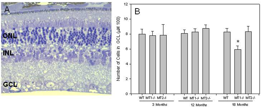
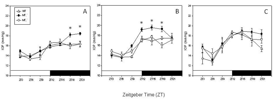
Previous studies have demonstrated that melatonin is effective in lowering intraocular pressure and that it may also protect ganglion cells. We have recently reported that, in mice lacking the melatonin receptors type 1, 25-30% ganglion cells die out by 18months of age, suggesting that these receptors might be important for ganglion cells survival. In this study we show that the loss of ganglion cells is specific for melatonin receptors type 1 knock-out since mice lacking the melatonin receptors type 2 did not show any significant change in the number ganglion cells during aging. Furthermore, we report that melatonin receptors type 1 knock-out mice have higher intraocular pressure during the nocturnal hours than control or melatonin receptors type 2 knock-out mice at 3 and 12months of age. Finally, our data indicate that administration of exogenous melatonin in wild-type, but not in melatonin receptors type 1 knock-out, can significantly reduce intraocular pressure. Our studies indicate that the decreased viability of ganglion cells observed in melatonin receptors type 1 knock-out mice may be a consequence of the increases in the nocturnal intraocular pressure thus suggesting that intraocular pressure levels at night and melatonin signaling should be considered as risk factor in the pathogenesis of glaucoma.
Copyright © 2011 Elsevier Ireland Ltd. All rights reserved.
Figures

References
-
- Aihara M, Lindsey JD, Weinreb RN. Twenty-four-hour pattern of mouse intraocular pressure. Exp. Eye Res. 2003;77:681–686. 2003. - PubMed
-
- Alarma-Estrany P, Pintor J. Melatonin receptors in the eye: location, second messengers and role in ocular physiology. Pharmacol. Ther. 2007;113:507–522. - PubMed
-
- Alarma-Estrany P, Crooke A, Mediero A, Peláez T, Pintor J. Sympathetic nervous system modulates the ocular hypotensive action of MT2-melatonin receptors in normotensive rabbits. J. Pineal Res. 2008;45:468–475. - PubMed
-
- Alarma-Estrany P, Crooke A, Pintor J. 5-MCA-NAT does not act through NQO2 to reduce intraocular pressure in New-Zealand white rabbit. J Pineal Res. 2009;47:201–219. - PubMed
-
- Baba K, Pozdeyev N, Mazzoni F, Contreras-Alcantara S, Liu C, Kasamatsu M, Martinez-Merlos T, Strettoi E, Iuvone PM, Tosini G. Melatonin modulates visual function and cell viability in the mouse retina via the MT1 melatonin receptor. Proc. Natl. Acad. Sci. U.S. A. 2009;106:15043–15048. - PMC - PubMed
Publication types
MeSH terms
Substances
Grants and funding
LinkOut - more resources
Full Text Sources
Molecular Biology Databases
