. 2011 May 15;50(10):1368-81.
doi: 10.1016/j.freeradbiomed.2011.02.021. Epub 2011 Mar 11.

Cannabidiol protects against hepatic ischemia/reperfusion injury by attenuating inflammatory signaling and response, oxidative/nitrative stress, and cell death

Affiliations

Cannabidiol protects against hepatic ischemia/reperfusion injury by attenuating inflammatory signaling and response, oxidative/nitrative stress, and cell death

Partha Mukhopadhyay et al. Free Radic Biol Med. .

Abstract

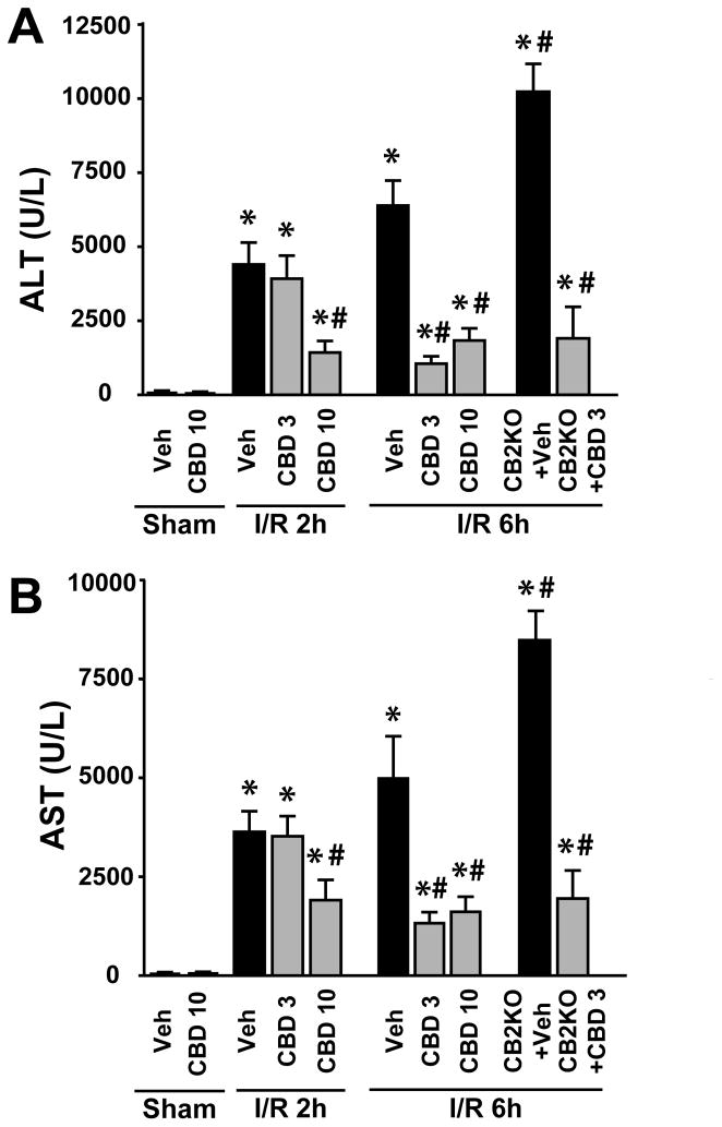
Ischemia/reperfusion (I/R) is a pivotal mechanism of liver damage after liver transplantation or hepatic surgery. We have investigated the effects of cannabidiol (CBD), the nonpsychotropic constituent of marijuana, in a mouse model of hepatic I/R injury. I/R triggered time-dependent increases/changes in markers of liver injury (serum transaminases), hepatic oxidative/nitrative stress (4-hydroxy-2-nonenal, nitrotyrosine content/staining, and gp91phox and inducible nitric oxide synthase mRNA), mitochondrial dysfunction (decreased complex I activity), inflammation (tumor necrosis factor α (TNF-α), cyclooxygenase 2, macrophage inflammatory protein-1α/2, intercellular adhesion molecule 1 mRNA levels; tissue neutrophil infiltration; nuclear factor κB (NF-κB) activation), stress signaling (p38MAPK and JNK), and cell death (DNA fragmentation, PARP activity, and TUNEL). CBD significantly reduced the extent of liver inflammation, oxidative/nitrative stress, and cell death and also attenuated the bacterial endotoxin-triggered NF-κB activation and TNF-α production in isolated Kupffer cells, likewise the adhesion molecule expression in primary human liver sinusoidal endothelial cells stimulated with TNF-α and attachment of human neutrophils to the activated endothelium. These protective effects were preserved in CB2 knockout mice and were not prevented by CB1/2 antagonists in vitro. Thus, CBD may represent a novel, protective strategy against I/R injury by attenuating key inflammatory pathways and oxidative/nitrative tissue injury, independent of classical CB1/2 receptors.


Conflict of interest statement

Disclosures: No conflict of interest to disclose.

Figures

Figure 1
Figure 1. Cannabidiol pretreatment decreases liver I/R injury
Panel A-B: Serum transaminase ALT (A) and AST (B) levels in sham operated mice treated with vehicle (veh) or CBD (n=5/group) or in mice exposed to 1 h of hepatic ischemia followed by 2 or 6 hours of reperfusion pretreated with vehicle or CBD (3 or 10 mg/kg, n=7–12/group). *P<0.05 vs. vehicle-sham group; #P<0.05 vs. corresponding control or cannabinoid 2 receptor knockout (CB2KO) mice exposed to vehicle-I/R.
Figure 2
Figure 2. Cannabidiol treatment after ischemia or at 90 mins of reperfusion decreases liver I/R injury
Panel A and B: Serum transaminase ALT and AST levels in mice exposed to 1 h of hepatic ischemia followed by 6 hours of reperfusion treated with vehicle or CBD (3 or 10 mg/kg, n=5–7/group) either right after the ischemia before reperfusion (panel A) or at 90 minutes of reperfusion (panel B). #P<0.05 vs. corresponding mice exposed to vehicle-I/R.
Figure 3
Figure 3. Cannabidiol decreases histological damage 24h following ischemia
Hematoxylin and eosin staining of representative liver sections of sham mice treated with vehicle (sham) or CBD (CBD), and mice exposed to 1 hour of ischemia followed by 24 hours of reperfusion, treated with vehicle (I/R) or CBD (I/R+CBD). A similar histological profile was seen in three to five livers/group. Upper row of images depicts 200x magnification, while the lower one 400x magnification.
Figure 4
Figure 4. Cannabidiol attenuates I/R-induced enhanced hepatic cell death
Panels A: Representative TUNEL staining 24 h following I/R injury. TUNEL positive nuclei of cells are light blue/white (colocalization of TUNEL (green staining) with nuclear staining (blue)), whereas the dark blue staining represents the staining of nuclei of normal cells. Right panel: quantification of hepatic TUNEL staining. Results are mean±SEM of 10–12 frames/group from 3–4 different animals/group. *P<0.05 vs. sham vehicle; #P<0.05 vs. I/R vehicle mice. Panels B: Time-dependent increase of hepatic DNA fragmentation demonstrated following I/R injury and attenuation by CBD pretreatment at 10 mg/kg. Results are mean±SEM of n=8/group. *P<0.05 vs. sham vehicle; #P<0.05 vs. corresponding I/R mice. Panels C: Time dependent changes in hepatic PARP activity following I/R, and attenuation of the observed increases by pretreatment with CBD at 10 mg/kg. Results are mean±SEM of n=8/group. *P<0.05 vs. sham vehicle; #P<0.05 vs. corresponding I/R mice.
Figure 5
Figure 5. Cannabidiol attenuates I/R-induced acute pro-inflammatory chemokines response in the liver
Panels A: Real-time PCR shows significant increase of chemokine MIP-1α (CCL3) mRNA level at 2 hours of reperfusion (IR 2h), and a decrease at 24 hours of reperfusion (IR 24h). Pretreatment with CBD at 10 mg/kg significantly attenuates the I/R-induced increased pro-inflammatory chemokine levels at all time points of the reperfusion studied (2, 6 and 24 hours). Panels B: Real-time PCR shows significant increase of chemokine MIP-2 (CXCL2) mRNA level at 2 h of reperfusion (IR 2h), and a decrease at 24 hours of reperfusion (IR 24h). Pretreatment with CBD at 10 mg/kg attenuates the I/R-induced increased pro-inflammatory chemokine levels at all time points of the reperfusion studied (2, 6 and 24 hours). Results are mean±SEM of 6–12 mice/groups. *P<0.05 vs. vehicle-sham group; #P<0.05 vs. corresponding vehicle-I/R mice.
Figure 6
Figure 6. Cannabidiol attenuates I/R-induced increased adhesion molecule expression and acute pro-inflammatory cytokine TNF-α response in the liver
Panels A: Real-time PCR shows significant increase of hepatic adhesion molecule ICAM-1 mRNA level at 2 hours of reperfusion (IR 2h), which was further attenuated at 6 and 24 hours of reperfusion (I/R 6h and 24h). Pretreatment with CBD at 10 mg/kg significantly attenuates the I/R-induced increased hepatic ICAM-1 expression at all time points of the reperfusion studied (2, 6 and 24 hours). Panels B: Real-time PCR shows significant increase of hepatic pro-inflammatory cytokine TNF-α mRNA level at 2 h of reperfusion (IR 2h), and a gradual decrease with time. Pretreatment with CBD at 10 mg/kg attenuates the I/R-induced increased hepatic TNF-α mRNA level at all time points of reperfusion studied (2, 6 and 24 hours). Results are mean±SEM of 6–12 mice/groups. *P<0.05 vs. vehicle-sham group; #P<0.05 vs. corresponding vehicle-I/R mice.
Figure 7
Figure 7. Cannabidiol decreases I/R-induced neutrophil infiltration after I/R injury
Myeloperoxidase staining (brown) of representative liver sections of sham mice pretreated with vehicle (sham) or CBD (CBD), and mice exposed to 1 hour of hepatic ischemia followed by 24 hours of reperfusion with vehicle (I/R) or CBD (I/R+CBD) pretreatment. I/R followed by 24 hours of reperfusion dramatically increased neutrophil infiltrationin in the livers, which was attenuated by CBD pretreatment. In livers of sham operated mice with or without pretreatment there was no tissue inflammatory cell infiltration, likewise only a very few inflammatory cells were present in the lumen of some vessels at 2 hours and 6 of reperfusion. Slides were counterstained by nuclear fast red. A similar histological profile was seen in three to five livers/group. Upper row of images depicts 200x magnification, while the lower one 400x magnification.
Figure 8
Figure 8. Cannabidiol attenuates I/R-induced hepatic NF-κB activation and p38 MAPK and JNK phosphorylation
Panel A: The gel shift assay demonstrates NF-κB activation at 2 and 6 hours of reperfusion (I/R 2h and I/R 6h) which was attenuated at 24 hours reperfusion (I/R 24h). CBD pretreatment at 10 mg/kg attenuates these activation at all time points of reperfusion studied. Panel B: Marked increase in the p38MAPK and c-Jun N-terminal kinase (JNK) activation in I/R liver tissues of mice at 2 and 6 hours following I/R injury, which are attenuated by CBD pretreatment at 10 mg/kg. β-actin is used as loading control.
Figure 9
Figure 9. Cannabidiol decreases the I/R-induced increased hepatic oxidative stress and restores decreased mitochondrial complex I activity
Panel A: HNE adduct, a marker for lipid peroxidation/oxidative stress, increases with time following I/R injury, and CBD pretreatment at 10 mg/kg attenuates these increases at all time points of reperfusion studied (2, 6 and 24 hours). Results are mean±SEM of 8 mice/groups. *P<0.05 vs. vehicle-sham group; #P<0.05 vs. corresponding vehicle-I/R mice. Panel B: Mitochondrial complex I activity decreased at 2 hours of reperfusion which partially recovered at 6 and 24 hours of reperfusion. Pretreatment with CBD at 10 mg/kg attenuated the I/R-induced decreased mitochondrial complex I at all time points of the reperfusion studied (2, 6 and 24 hours). Results are mean±SEM of 6–7 mice/groups. *P<0.05 vs. vehicle-sham group; #P<0.05 vs. corresponding vehicle-I/R mice. Panel C: Real-time PCR shows significant increase of hepatic gp91phox mRNA level from 6 hours of reperfusion (IR 6h), which peaks at 24 hours (IR 24h). Pretreatment with CBD at 10 mg/kg significantly attenuates the I/R-induced increased liver gp91phox mRNA expression at 6 and 24 hours of reperfusion. Results are mean±SEM of 8–10 mice/groups. *P<0.05 vs. vehicle-sham group; #P<0.05 vs. corresponding vehicle-I/R mice.
Figure 10
Figure 10. Cannabidiol attenuates I/R-induced COX-2 and iNOS mRNA expression and nitrative stress in the liver
Panel A: Real-time PCR shows significant time-dependent increases of hepatic COX-2 mRNA level at 2, 6 and 24 hours of reperfusion (IR 2h, 6h and 24h). Pretreatment with CBD at 10 mg/kg attenuates the I/R-induced increase in COX-2 mRNA expression at all time points of the reperfusion studied (2, 6 and 24 hours). Results are mean±SEM of 6–10 mice/groups. *P<0.05 vs. vehicle-sham group; #P<0.05 vs. corresponding vehicle-I/R mice. Panel B: Real-time PCR shows significant time-dependent increases of hepatic iNOS mRNA level at 2, 6 and 24 hours of reperfusion (IR 2h, 6h and 24h). Pretreatment with CBD at 10 mg/kg attenuates the I/R-induced increase in iNOS mRNA at all time points of the reperfusion studied (2, 6 and 24 hours). Results are mean±SEM of 6–10 mice/groups. *P<0.05 vs. vehicle-sham group; #P<0.05 vs. corresponding vehicle-I/R mice. Panels C: Nitrotyrosine modification of protein, a marker for nitrative stress, increases with time following I/R injury, and CBD pretreatment at 10 mg/kg attenuates these increases at all time points of reperfusion studied (2, 6 and 24 hours). Results are mean±SEM for both panels and n=8–12/group. *P<0.05 vs. vehicle; #P<0.05 vs. corresponding vehicle-I/R mice.
Figure 11
Figure 11. Cannabidiol attenuates I/R-induced hepatic nitrotyrosine staining
Panel A: Increased nitrotyrosine staining after 6 hours of liver reperfusion in perivascular area (endothelial cells and surrounding layers of hepatocytes), which was attenuated by CBD treatment (200x magnification). Similar pattern, but weaker staining was seen at 2 hours of reperfusion (not shown). Panel B: Very strong hepatic nitrotyrosine staining in perivascular area at 24 hours of reperfusion (200x magnification). On the magnified insert (middle image) it is clearly shown that the strong nitrotyrosine staining was present in hepatocytes around the vessels, endothelial cells, and in inflammatory infiltrate (neutrophil granulocytes, macrophages, etc.). CBD was able to attenuate the I/R-induced nitrotyrosine formation. Panel C shows further details of nitrotyrosine staining after 24 hours of reperfusion with a 1000x magnification in endothelial cells, hepatocytes and in the inflammatory infiltrate. Similar staining was seen in 4 to 6 livers/group.
Figure 12
Figure 12. Cannabidiol attenuates LPS-induced NF-κB activation and TNFα secretion in Kupffer cells
Panel A: Western blot analysis demonstrates inhibition of nuclear transcription factor NF-κB (IκB) expression and its phosphorylation in the cytosolic fraction. CBD attenuates LPS induced phosphorylation of IκB in Kupffer cells. β-Actin is used as loading control. Panel B: CBD (0.3–3 μM) attenuates LPS induced TNFα secretion of Kupffer cells in dose-dependent manner. Results are mean±SEM of n=4–6/group. *P<0.05 vs. vehicle; #P<0.05 vs. LPS.
Figure 13
Figure 13. Cannabidiol attenuates the TNF-α induced adhesion molecules expression in human liver sinusoidal endothelial cells (HLSEC)
Treatment of HLSEC cells with TNF-α for 6 hrs markedly enhances the expression of adhesion molecules such as ICAM-1 and VCAM-1. CBD (1 μM) attenuates this enhanced expression of adhesion molecules ICAM-1 (Panel A) and VCAM-1 (Panel B). Cannabinoid receptors antagonists/inverse agonists such as SR141716 (SR1 for CB1 receptor) and SR144528 (SR2 for CB2 receptor) at 1 μM could not prevent the CBD-mediated attenuation of TNF-α-induced adhesion molecules expression. Results are mean±SEM of n=4–6/group. *P<0.05 vs. vehicle; #P<0.05 vs. TNF-α.
Figure 14
Figure 14. Cannabidiol attenuates the TNF-α induced polymorphonuclear cells (PMN) adhesion to human liver sinusoidal endothelial cells (HLSEC)
Representative images and quantification of human neutrophil adhesion to activated human liver endothelial cells. Treatment of HLSEC cells with TNF-α (50 ng/ml) for 6 hrs markedly enhances PMN adhesion. CBD (1 μM) attenuates this enhanced PMN adhesion. Cannabinoid receptors antagonists/inverse agonists such as SR141716 (SR1 for CB1 receptor) and SR144528 (SR2 for CB2 receptor) at 1 μM could not prevent the CBD-mediated attenuation of TNF-α-induced PMN adhesion. Results are mean±SEM of n=4–6/group. *P<0.05 vs. vehicle; #P<0.05 vs. TNF-α.

Similar articles

Cited by

References

    1. Liaudet L, Szabo G, Szabo C. Oxidative stress and regional ischemia-reperfusion injury: the peroxynitrite-poly(ADP-ribose) polymerase connection. Coron Artery Dis. 2003;14:115–122. - PubMed
    1. Ferdinandy P, Schulz R. Nitric oxide, superoxide, and peroxynitrite in myocardial ischaemia-reperfusion injury and preconditioning. Br J Pharmacol. 2003;138:532–543. - PMC - PubMed
    1. Pacher P, Beckman JS, Liaudet L. Nitric oxide and peroxynitrite in health and disease. Physiol Rev. 2007;87:315–424. - PMC - PubMed
    1. Hines IN, Grisham MB. Divergent roles of superoxide and nitric oxide in liver ischemia and reperfusion injury. J Clin Biochem Nutr. 2011;48:50–56. - PMC - PubMed
    1. Szabo C. The pathophysiological role of peroxynitrite in shock, inflammation, and ischemia-reperfusion injury. Shock. 1996;6:79–88. - PubMed

Publication types