Review
. 2011 Feb;38(1):3-15.
doi: 10.1053/j.seminoncol.2010.11.010.

The evolution of imaging in cancer: current state and future challenges

Affiliations
Review

The evolution of imaging in cancer: current state and future challenges

Luke J Higgins et al. Semin Oncol. 2011 Feb.

Abstract

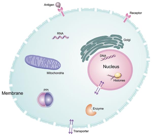
Molecular imaging allows for the remote, noninvasive sensing and measurement of cellular and molecular processes in living subjects. Drawing upon a variety of modalities, molecular imaging provides a window into the biology of cancer from the subcellular level to the patient undergoing a new, experimental therapy. As signal transduction cascades and protein interaction networks become clarified, an increasing number of relevant targets for cancer therapy--and imaging--become available. Although conventional imaging is already critical to the management of patients with cancer, molecular imaging will provide even more relevant information, such as early detection of changes with therapy, identification of patient-specific cellular and metabolic abnormalities, and the disposition of therapeutic, gene-tagged cells throughout the body--all of which will have a considerable impact on morbidity and mortality. This overview discusses molecular imaging in oncology, providing examples from a variety of modalities, with an emphasis on emerging techniques for translational imaging.


Figures

Figure 1
Figure 1
Some cellular targets for molecular imaging. Oncologic molecular imaging agents exist for each of these targets. In addition to standard imaging agent binding to extracellular targets in the membrane, eg, G-protein–coupled receptors, some agents localize to various organelles, such as lysosomes, mitochondria or golgi apparatus, based on electrostatic potential or pH. PPI , protein–protein interactions, as detected by split luciferase reporters.
Figure 2
Figure 2
Lysine-rich protein (LRP) as an MR-CEST reporter. (Top) Illustration of the chemical exchange saturation transfer (CEST) contrast mechanism. A frequency-selective radiofrequency pulse is used to label (green) the amide protons of the contrast agent. Only labeled protons exchange with water protons. This leads to a reduction in MR signal intensity. (Bottom) CEST difference map (right) overlaid on an anatomical MR image (left). This map demonstrates that LRP-expressing xenografts can be distinguished from mock-transfected controls. Reprinted with permission from Macmillan Publishers Ltd: Nature Biotechnology, copyright 2007.
Figure 3
Figure 3
Case study: noninvasive detection of cytolytic T cells in a patient with glioma. MRI (top) and PET over MRI superimposed (bottom) brain images of a patient who received autologous cyolytic T-cell infusions. T cells expressed HSV1-TK and images were acquired after 18F-FHBG injection. Cells localized to tumor 1 and trafficked to tumor 2. Reprinted with permission from Macmillan Publishers Ltd: Nature Clin Pract Oncol, copyright 2007.
Figure 4
Figure 4
Case study: anti-[18F] FACBC to guide prostate cancer radiotherapy target design. A CT image (A), an FACBC PET image (B), and a registered image (C) demonstrates how FACBC PET scan information was registered with the planning CT scan. The projection of FACBC-defined gross tumor volume (GTVFACBC) into the treatment planning CT at three different levels (D–F). GTVFACBC was mostly contained within the prostate. Reprinted with permission from Jani et al. Clin Nucl Med. 2009.

Similar articles

Cited by

References

    1. Ogawa S, Lee TM, Kay AR, Tank DW. Brain magnetic resonance imaging with contrast dependent on blood oxygenation. Proc Natl Acad Sci U S A. 1990;87:9868–72. - PMC - PubMed
    1. Patterson DM, Padhani AR, Collins DJ. Technology insight: water diffusion MRI—a potential new biomarker of response to cancer therapy. Nat Clin Pract Oncol. 2008;5:220–33. - PubMed
    1. Lee KC, Bradley DA, Hussain M, et al. A feasibility study evaluating the functional diffusion map as a predictive imaging biomarker for detection of treatment response in a patient with metastatic prostate cancer to the bone. Neoplasia. 2007;9:1003–11. - PMC - PubMed
    1. Kwee TC, Takahara T, Klomp DW, Luijten PR. Cancer imaging: novel concepts in clinical magnetic resonance imaging. J Intern Med. 2010;268:120–32. - PubMed
    1. Kovacs A, Toth L, Glavak C, et al. Integrating functional MRI information into radiotherapy planning of CNS tumors—early experiences. Pathol Oncol Res. Epub ahead of print 2010 September 17. - PubMed

Substances