Comparative Study
. 2011 Mar;17(3):253-63.
doi: 10.1016/j.cardfail.2010.10.008. Epub 2010 Dec 24.

Left ventricular dysfunction in murine models of heart failure and in failing human heart is associated with a selective decrease in the expression of caveolin-3

Affiliations
Comparative Study

Left ventricular dysfunction in murine models of heart failure and in failing human heart is associated with a selective decrease in the expression of caveolin-3

Ellina Cheskis Feiner et al. J Card Fail. 2011 Mar.

Abstract

Background: Caveolins are scaffolding proteins that are integral components of caveolae, flask-shaped invaginations in the membranes of all mammalian cells. Caveolin-1 and -2 are expressed ubiquitously, whereas caveolin-3 is found only in muscle. The role of caveolin-3 in heart muscle disease is controversial.

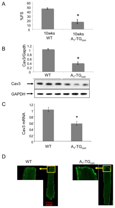
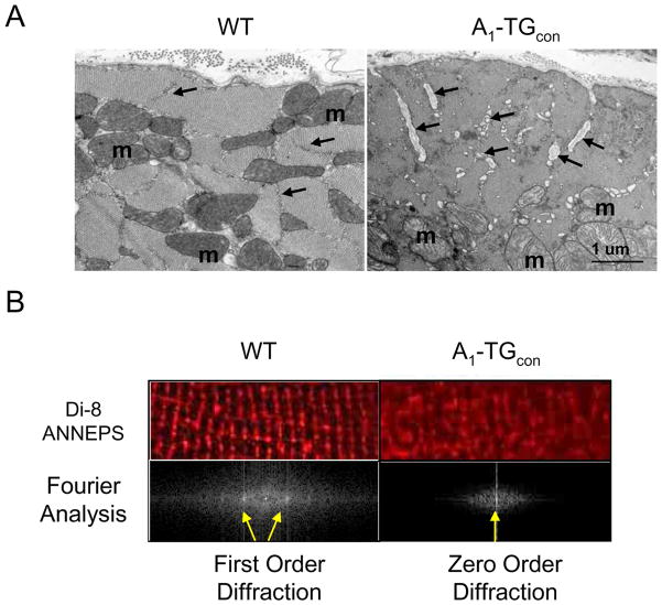
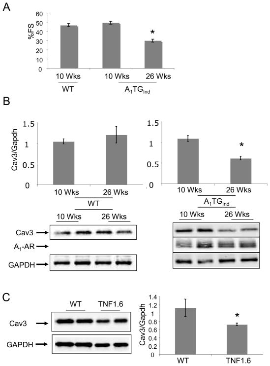
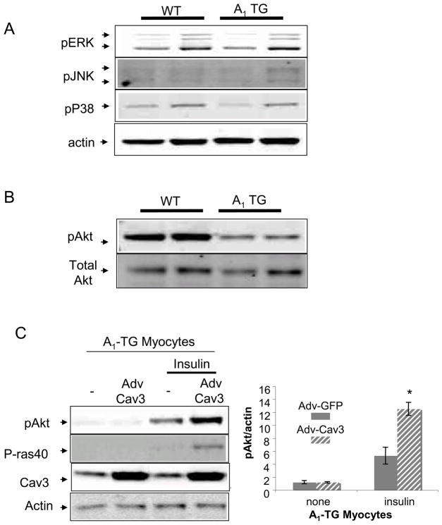
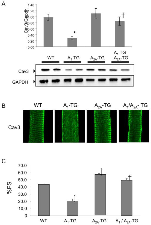
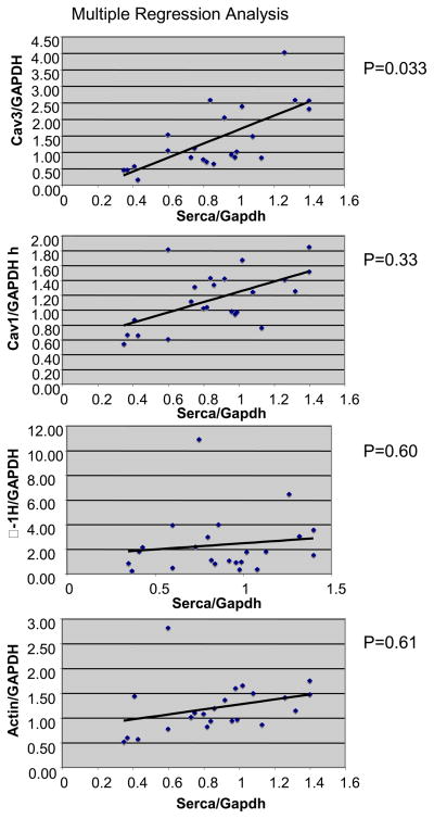
Methods and results: The present study was undertaken to assess the effects of left ventricular dysfunction on the expression of caveolin proteins using 2 well characterized models of murine heart failure and failing human heart. Transgenic mice with constitutive overexpression of A(1)-adenosine receptor (A(1)-TG) demonstrated cardiac dilatation and decreased left ventricular function at 10 weeks of age. This was accompanied by a marked decrease in caveolin-3 mRNA and protein levels compared with non-TG control mice. The change in caveolin-3 expression was selective, because levels of caveolin-1 and -2 did not change. Confocal imaging of myocytes isolated from A(1)-TG mice demonstrated a loss of the plate-like appearance of T tubules. Caveolin-3 levels were also reduced in hearts from mice overexpressing tumor necrosis factor α. There was a direct relationship between caveolin-3 expression and fractional shortening in all mice that were studied (r = 0.65; P < .001). Although we could not demonstrate a significant decrease in caveolin-3 levels in failing human heart, we did find a direct correlation (r = 0.7; P < .05) between levels of caveolin-3 protein and Ca(2+)-adenosine triphosphatase, a marker of the heart failure phenotype.

Conclusions: These results suggest a relationship between left ventricular dysfunction and caveolin-3 levels and suggest that caveolin-3 may provide a novel target for heart failure therapy.


Figures

Figure 1
Figure 1
(A) Fractional shortening percentage (%FS) measurement by echocardiograph. 10 week-old WT and A1-TGcon mice were used (N=5-8). *P<0.01. (B) Caveolin-3 protein expression. Ventricular extracts from 10 week-old mice were probed with indicated antibodies. Signals were normalized to GAPDH expression in WT hearts (N=5) and were analyzed by non-parametric method. Analyzed values were mean+/−SE, *P<0.05 vs WT. (C) Reverse-transcribed total RNA from mouse left ventricles were used to detect caveolin-3 and GAPDH gene expression. Signals were normalized to GAPDH expression in WT hearts (N=5) and were analyzed by non-parametric method. Analyzed values were mean+/−SE, *P<0.05 vs WT. (D) Immunofluorescence caveolin-3 staining of isolated myocytes. 600X confocal images represent at least 3 mice/genotype group and minimum 20 myocytes/mice examined. myocytes pooled from 4-5 mice/genotype group.
Figure 2
Figure 2
A. Electron microscopy images of myocardial sections of male 12–14 week WT (n=3) and A1-TGcon (n=3) mouse hearts. T-tubules (arrows) are dilated in the transgenic cardiac muscle. The dilated T tubules contain membranous and basement membrane-like material; m=mitochondrion. Bar = 1 micron. B. T-tubules were stained with the voltage sensitive lipid dye, di-8-ANNEPS, in live WT and A1-TGcon myocytes. Confocal digital images were processed using Fast Fourier Transform (FFT) algorithm to quantify organization of T-tubule within myocytes. The power spectrum in log scale vs spatial frequency was calculated for each image and indicated by the appearance of zero or first order diffraction spots (arrows).
Figure 3
Figure 3
(A) Adult A1-AR induction. Doxycycline (DOX) was removed from transgenic mice at 3 weeks of age to induce A1-AR expression (A1-TGInd). Percent fractional shortening (%FS) were determined in A1-TGInd mice at 10 week-of-age and at 26 week-of-age. *P<0.05 vs 10 week-old A1-TGInd. (B) Caveolin-3, A1-AR and GAPDH expression in 10–12 week-old WT (N=8), 20–26 week-old WT (N=4), 10–12 week-old A1-TGind (N=4) and 20–26 week-old A1-TGind (N=8) mouse left ventricles. Data shown are mean +/− SE. *p<0.05. (C) Caveolin-3 and GAPDH expression in 12 week-old WT and TNF 1.6 left ventricular tissues (N=4). Data shown are mean +/− SE. *p<0.05. (D) Cardiac function (FS%) and caveolin-3 protein expression were obtained from mice with the following genotypes: WT FVB mice (N=4), 10 wk-old A1-TGcon (N=6), 10 wk-old A1-TGInd (N=8), 26 wk-old A1-TGInd (N=6), 10 wk-old A1/A2A-TGcon (N=5), TNF 1.6 (N=4). Non-parametric Spearman Rank test was used to correlate Cav3 expression with fractional shortening. Graph showed FS% and Caveolin-3/GAPDH scatter plot. Linear trend bar showed the significant correlation between caveolin-3 expression and fractional shortening (r=0.65; p<0.001).
Figure 3
Figure 3
(A) Adult A1-AR induction. Doxycycline (DOX) was removed from transgenic mice at 3 weeks of age to induce A1-AR expression (A1-TGInd). Percent fractional shortening (%FS) were determined in A1-TGInd mice at 10 week-of-age and at 26 week-of-age. *P<0.05 vs 10 week-old A1-TGInd. (B) Caveolin-3, A1-AR and GAPDH expression in 10–12 week-old WT (N=8), 20–26 week-old WT (N=4), 10–12 week-old A1-TGind (N=4) and 20–26 week-old A1-TGind (N=8) mouse left ventricles. Data shown are mean +/− SE. *p<0.05. (C) Caveolin-3 and GAPDH expression in 12 week-old WT and TNF 1.6 left ventricular tissues (N=4). Data shown are mean +/− SE. *p<0.05. (D) Cardiac function (FS%) and caveolin-3 protein expression were obtained from mice with the following genotypes: WT FVB mice (N=4), 10 wk-old A1-TGcon (N=6), 10 wk-old A1-TGInd (N=8), 26 wk-old A1-TGInd (N=6), 10 wk-old A1/A2A-TGcon (N=5), TNF 1.6 (N=4). Non-parametric Spearman Rank test was used to correlate Cav3 expression with fractional shortening. Graph showed FS% and Caveolin-3/GAPDH scatter plot. Linear trend bar showed the significant correlation between caveolin-3 expression and fractional shortening (r=0.65; p<0.001).
Figure 4
Figure 4
Signal transduction in A1-TG hearts. (A) Extracts of 10 week-old WT and A1-TG mouse left ventricles were probed with antibodies specific for Phospho-ERK, phospho-JNK and phospho-p38. (B) Extracts of 10 week-old WT and A1-TG mouse left ventricles were probed with antibodies specific for Phospho-Akt and total Akt. (C) Adv-Caveolin-3 expression in A1-TG myocytes. Adult myocytes were isolated from 20-week-old adult induced A1-TG mice. After infecting cells with Adv-Caveolin-3 virus or control Adv-GFP virus, myocytes were cultured for 48hrs. Then, cells were acutely stimulated with serum-free medium containing the Insulin-Transferrin-Selenium supplement (Invitrogen Corporation) for 15 minutes and probed with indicated antibodies. Blots shown were representative of at least three independent experiments. The graph showed pAkt/actin ratio: mean +/− SE. *p<0.05.
Figure 5
Figure 5
Caveolin (Cav-3) protein expression, caveolin-3 localization and cardiac function in transgenic mice overexpressing both A1-R and A2A-R. (A) caveolin-3 expression in WT, A1-TG, A2A-TG and A1/A2A-TG mice. Ventricular extracts from 10-week-old age and sex matched mice (N=4) were probed with indicated antibodies. Signals were normalized to GAPDH expression in WT hearts and were analyzed by non-parametric method. Analyzed values were mean+/-SE, *P<0.05 vs WT. (B) Caveolin-3 localization in cardiac myocytes. 600X confocal images are shown. (C) Percent fractional shortening (%FS) of indicated mouse groups. *P<0.001 vs WT, P<0.001 vs A1TG. (n=15–21, SE, male 8–12 week old mice).
Figure 6
Figure 6
Correlation of SERCA expression with caveolin-1, caveolin-3, α1-H and actin expression in human failing hearts. Caveolin-1, caveolin-3, SERCA, α1-H and actin expression were measured in 5 normal human hearts and 19 hearts from heart failure patients. GAPDH-normalized SERCA expression in each heart was compared to the expression of other proteins by multiple regression analysis. Scatter plots with correlation trend bars indicated the following p values: Serca vs Caveolin-3, p<0.05. Serca vs Caveolin-1, p=0.34; Serca vs α1-H, p=0.60; Serca vs actin, p=0.61.
Figure 7
Figure 7
Fractional shortening percentage (%FS) measurements of GiCT-TGind, A1-TGind, and A1/GiCT- TGind mice at 26 weeks-of-age, N=5 per group. Values are means +/− SE. *P<0.05 versus A1-TGind.

References

    1. Isshiki MAR. Calcium signal transduction from caveolae. Cell Calcium. 1999;26:201–8. - PubMed
    1. Isshiki MAJ, Yamamoto K, Fujita T, Ying Y, Anderson RGW. Sites of Ca(2+) wave initiation move with caveolae to the trailing edge of migrating cells. J Cell Sci. 2002;115:475–84. - PubMed
    1. Fujimoto T. Calcium pump of the plasma membrane is localized in caveolae. J Cell Biol. 1993;120:1147–57. - PMC - PubMed
    1. Lasley RDNP, Uittenbogaard A, Smart EJ. Activated cardiac adenosine A1 receptors translocate out of caveolae. J Biol Chem. 2000;275:4417–21. - PubMed
    1. Escriche MBJ, Ciruela F, Canela EI, Mallol J, Enrich C, Lluis C, Franco R. Ligand-induced caveolae-mediated internalization of A1 adenosine receptors: morphological evidence of endosomal sorting and receptor recycling. Experimental Cell Research. 2003;285:72–90. - PubMed

Publication types