. 2011 Mar 4;41(5):567-78.
doi: 10.1016/j.molcel.2011.01.026.

Structure of lipid kinase p110β/p85β elucidates an unusual SH2-domain-mediated inhibitory mechanism

Affiliations

Structure of lipid kinase p110β/p85β elucidates an unusual SH2-domain-mediated inhibitory mechanism

Xuxiao Zhang et al. Mol Cell. .

Abstract

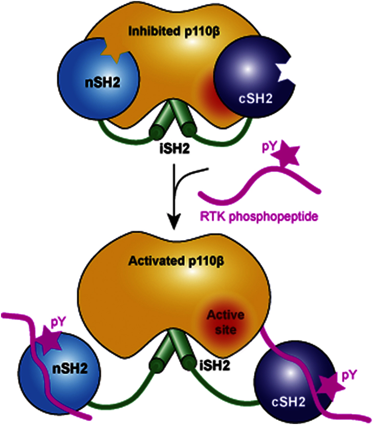
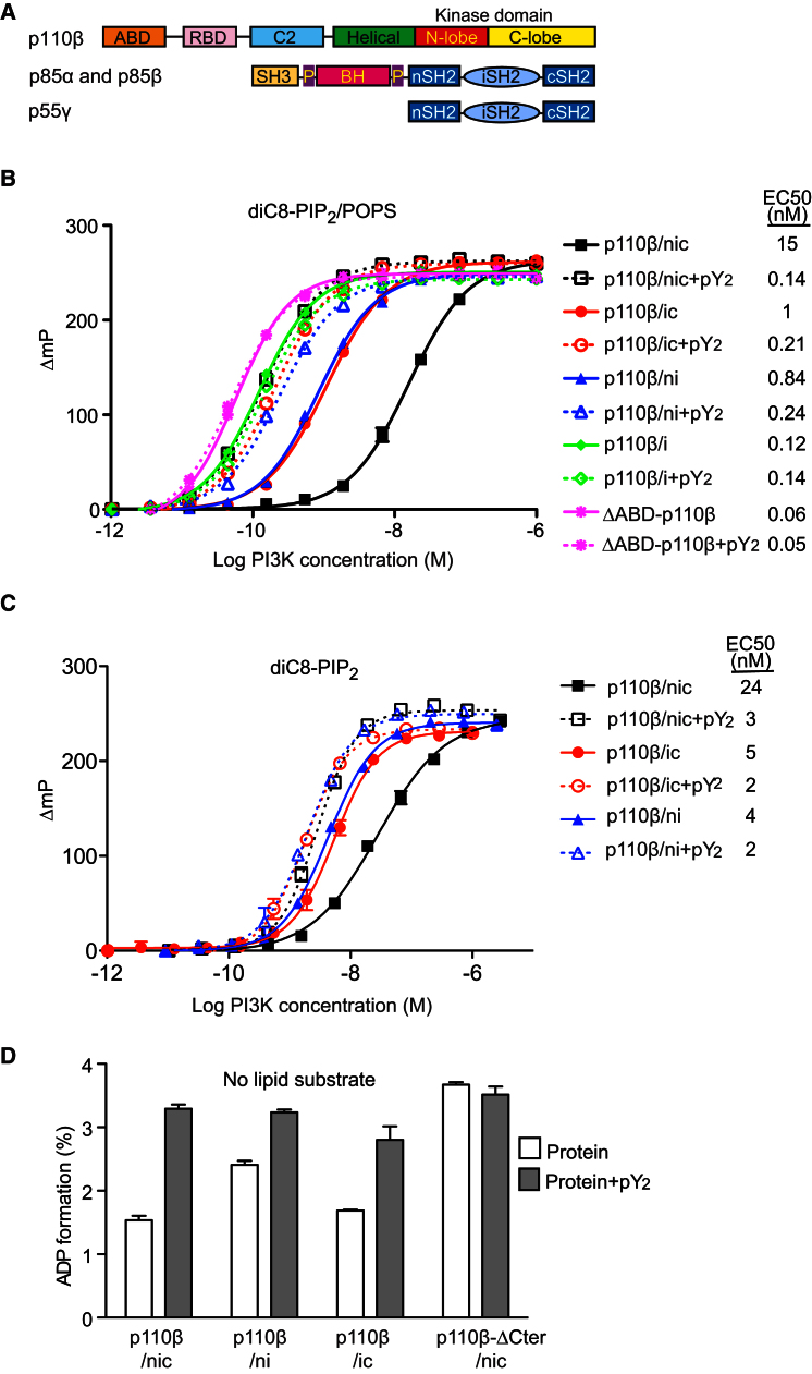
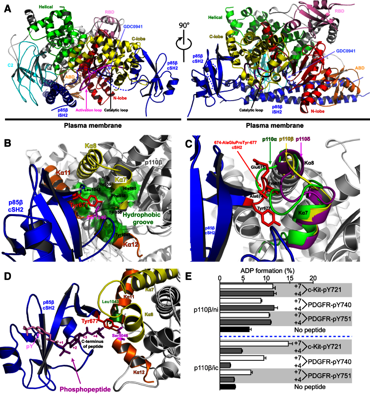
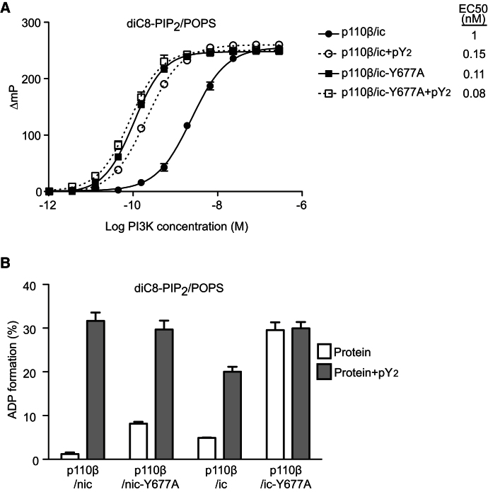
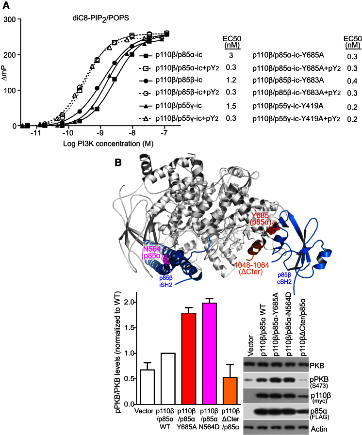
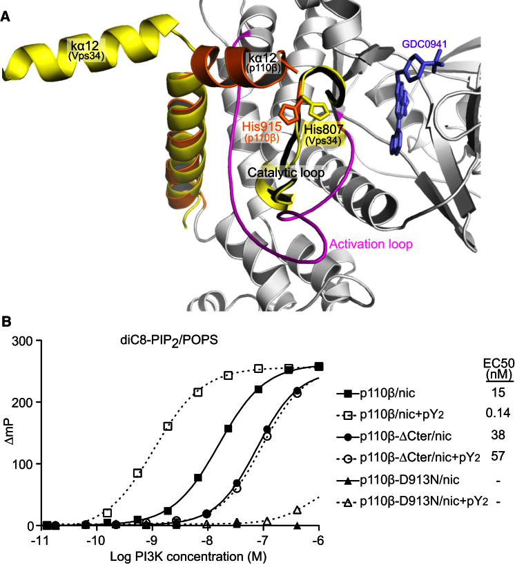
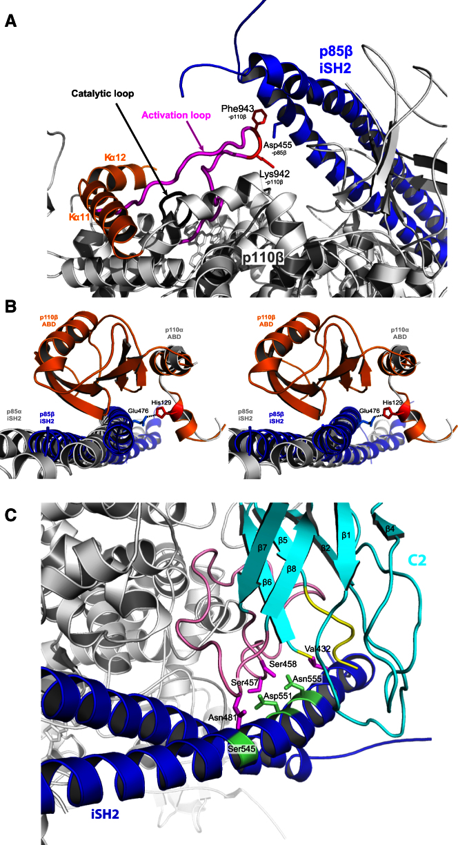
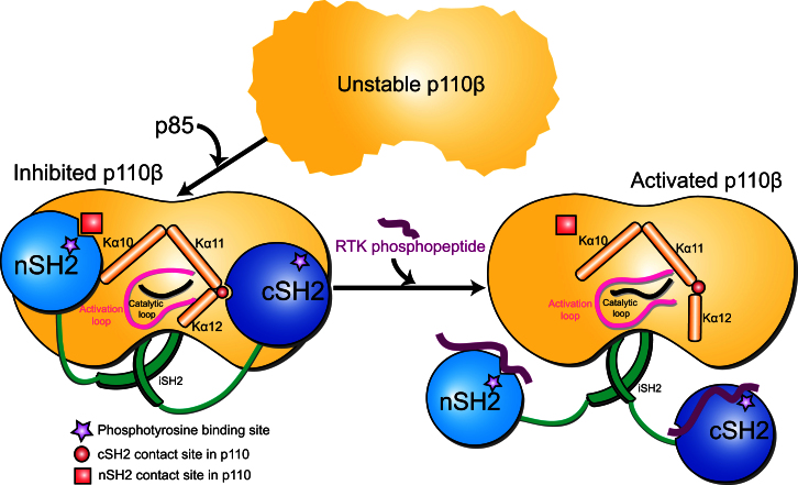
Phosphoinositide 3-kinases (PI3Ks) are essential for cell growth, migration, and survival. The structure of a p110β/p85β complex identifies an inhibitory function for the C-terminal SH2 domain (cSH2) of the p85 regulatory subunit. Mutagenesis of a cSH2 contact residue activates downstream signaling in cells. This inhibitory contact ties up the C-terminal region of the p110β catalytic subunit, which is essential for lipid kinase activity. In vitro, p110β basal activity is tightly restrained by contacts with three p85 domains: the cSH2, nSH2, and iSH2. RTK phosphopeptides relieve inhibition by nSH2 and cSH2 using completely different mechanisms. The binding site for the RTK's pYXXM motif is exposed on the cSH2, requiring an extended RTK motif to reach and disrupt the inhibitory contact with p110β. This contrasts with the nSH2 where the pY-binding site itself forms the inhibitory contact. This establishes an unusual mechanism by which p85 SH2 domains contribute to RTK signaling specificities.


Figures

None
Graphical abstract
Figure 1
Figure 1
Inhibition of p110β by p85-Type Regulatory Subunits and Activation of the Complexes by RTK Phosphopeptide (A) Domain organization of p110β and the regulatory subunits p85α, p85β, and p55γ. The color scheme for the domains is used for all figures, unless otherwise stated. (B) Kinase activity with diC8-PIP2/POPS liposomes as a function of enzyme concentration (measured by ADP formation) shows the inhibitory effects of p85β-nicSH2 (nic) and p85β-icSH2 (ic) on the basal activity of p110β. The inhibition is released upon addition of the 10 μM PDGFR pY2. The free catalytic subunit (ΔABD-p110β) is more active than any complex. The y axis is expressed as a change in fluorescence polarization (ΔmP), which is obtained by subtracting the observed polarization for the construct at a given enzyme concentration from the maximum fluorescence polarization for that construct. The plateau in these assays arises due to the competitive nature of the ADP detection system, based on displacement of ADP-Alexa 633 tracer from the ADP2-antibody by ADP generated during the PI3K assay (see Supplemental Experimental Procedures). All measurements were done in triplicates, and the error bars indicate the standard error of the mean (SEM). (C) Kinase activity of p110β/p85β complexes with monomeric substrate (75 μM diC8-PIP2) in the presence and absence of pY2; y axis as in (B). (D) Basal- and pY2-stimulated activities of 35 nM PI3K complexes in the absence of lipids (ATP hydrolysis). Activities are given as percent of ATP converted to ADP using the Transcreener assay. Bars indicate SEM.
Figure 2
Figure 2
Structure of p110β in Complex with p85β-icSH2 (A) Cartoon representation of the p110β/p85β-icSH2 complex with GDC0941 (light blue sticks). Views from the front and side (turned 90°) are shown. The ordered activation loop is colored in magenta and the catalytic loop in black. (B) Detailed view of the contacts between the p85β-cSH2 and the C-lobe of the kinase domain. The main contact residue Tyr677-p85β (red) interacts with Ser1046-p110β (magenta) and the hydrophobic groove (pale green) formed by two “arms,” Kα7/Kα8 (yellow) and Kα11/Kα12 (orange). Dashed black line is a potential hydrogen bond between Tyr677-p85β and Ser1046-p110β. (C) Cartoon of the p110β Kα7/Kα8 “arm” (yellow) superimposed on p110α (green) and p110δ (magenta), suggesting a steric clash between cSH2 protrusion (674-AlaGluPro-676) (red stick) and Kα7/Kα8 loop of p110α. (D) Cartoon of the cSH2 binding to RTK phosphopeptide, modeled as in the crystal structure of free cSH2 in complex with a PDGFR phosphopeptide (magenta; PDB ID: 1H9O). (E) Effects of different RTK phosphopeptides (10 μM) on activity of niSH2 and icSH2 complexes with p110β (1 nM), showing selective requirement for peptides longer than pY + 4 for disinhibition of the cSH2. Error bars indicate SEM.
Figure 3
Figure 3
Mutation Y677A in p85β-cSH2 Releases Inhibitory Effect on p110β (A) Kinase activity (ADP formation) of p110β in a complex with p85β-icSH2-Y677A (Y677A-ic) compared to the wild-type complex (ic) in the absence and presence of 10 μM PDGFR pY2 (ADP formation on the y axis expressed as in Figure 1B). (B) Comparison of p110β activities of 1 nM complexes with wild-type or Y677 mutant nicSH2 or icSH2 (activity shown as in Figure 1D).
Figure 4
Figure 4
cSH2 Is Important for p110β Inhibition In Vitro and in Cells (A) Kinase activity of human p110β and icSH2 wild-type constructs from three human regulatory subunits (p85α, p85β, and p55γ) in the absence and presence of 10 μM PDGFR pY2 (y axis as in Figure 1B). EC50s for wild-type and icSH2 tyrosine mutants, in the absence and presence of 10 μM PDGF pY2, are shown. (B) Western blots of HEK cells transiently expressing wild-type and mutant human p110β/p85α. The membrane was probed with antibodies against PKB, pPKB (pSer473), myc (p110β), FLAG (p85α), and actin. Bar graphs show mean ± SEM (n = 3) of PKB phosphorylation level normalized to wild-type p110β/p85α (WT). The position of the three mutations is mapped on the p110β/p85β structure.
Figure 5
Figure 5
Truncation of the C Terminus in p110β Decreases Its Activity for Lipid Substrates and Increases ATP Hydrolysis in the Absence of Lipids (A) The p110β kinase domain has the signatures of an inactive conformation: C-terminal Kα12 helix folds over the activation loop and His915 (from the DRH motif) points away from the active site (orange). They differ from the same elements in the presumably active conformation of Vps34 (yellow) (PDB: 2X6H). (B) Lipid kinase activity (ADP formation) of the wild-type p110β/p85α, in comparison with a truncation mutant lacking 17 residues from the p110β C terminus (ΔCter) and a kinase-dead mutant (D913N). Activities were determined in the absence and presence of 10 μM PDGFR pY2. For the D913N mutant, the EC50 was too high to be determined accurately (y axis as in Figure 1B).
Figure 6
Figure 6
Structural Basis for p110β Regulation by p85β-iSH2 Domain (A) Close-up of interactions between p85β-iSH2 (blue) and p110β activation loop. (B) Stereo view of the unique interaction between the iSH2 (blue) with the ABD-RBD linker of p110β (orange), as compared to p110α (white). (C) Detailed interactions between residues in the iSH2 (green sticks) with residues in the C2 domain of p110β (magenta sticks). The CBR3 (Cβ5/Cβ6 loop) and Cβ7/Cβ8 loops that contact the iSH2 are highlighted in yellow and pink, respectively.
Figure 7
Figure 7
A “Three-Brake” Regulatory Model for Inhibition of p110β Basal Activity Model of the “three-brake” inhibition of p110β activity by the nSH2, iSH2, and cSH2 domains of p85. All three domains make inhibitory interactions with the catalytic subunit. The nSH2 and cSH2 are in contact with the “regulatory square” formed by the three C-terminal helices of p110β (Kα10–Kα12). This “regulatory square” encloses the catalytic and activation loop and could mediate the SH2 inhibitory effects. RTK phosphopeptide binding to SH2 domains relieves the inhibition. The main contact between nSH2 and catalytic subunit overlaps perfectly with pY-binding site on nSH2, but not on cSH2. Therefore, relief from cSH2 inhibition requires an extended pYXXM motif.

Comment in

References

    1. Backer J.M., Myers M.G.J., Jr., Shoelson S.E., Chin D.J., Sun X.J., Miralpeix M., Hu P., Margolis B., Skolnik E.Y., Schlessinger J. Phosphatidylinositol 3′-kinase is activated by association with IRS-1 during insulin stimulation. EMBO J. 1992;11:3469–3479. - PMC - PubMed
    1. Bae J.H., Lew E.D., Yuzawa S., Tomé F., Lax I., Schlessinger J. The selectivity of receptor tyrosine kinase signaling is controlled by a secondary SH2 domain binding site. Cell. 2009;138:514–524. - PMC - PubMed
    1. Beeton C.A., Chance E.M., Foukas L.C., Shepherd P.R. Comparison of the kinetic properties of the lipid- and protein-kinase activities of the p110alpha and p110beta catalytic subunits of class-Ia phosphoinositide 3-kinases. Biochem. J. 2000;350:353–359. - PMC - PubMed
    1. Berndt A., Miller S., Williams O., Le D.D., Houseman B.T., Pacold J.I., Gorrec F., Hon W.C., Liu Y., Rommel C. The p110 delta structure: mechanisms for selectivity and potency of new PI(3)K inhibitors. Nat. Chem. Biol. 2010;6:117–124. - PMC - PubMed
    1. Bondeva T., Pirola L., Bulgarelli-Leva G., Rubio I., Wetzker R., Wymann M.P. Bifurcation of lipid and protein kinase signals of PI3Kgamma to the protein kinases PKB and MAPK. Science. 1998;282:293–296. - PubMed

Publication types

Substances

Associated data

LinkOut - more resources