. 2011 Mar 4;41(5):600-8.
doi: 10.1016/j.molcel.2011.02.004.

Coupled 5' nucleotide recognition and processivity in Xrn1-mediated mRNA decay

Affiliations

Coupled 5' nucleotide recognition and processivity in Xrn1-mediated mRNA decay

Martin Jinek et al. Mol Cell. .

Abstract

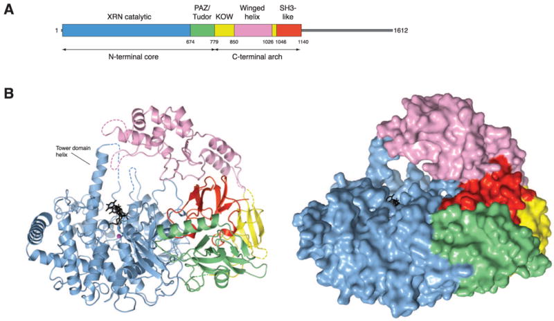
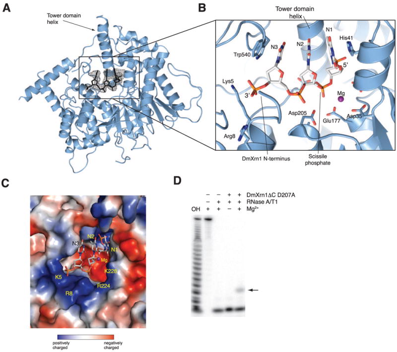
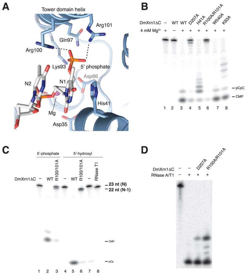
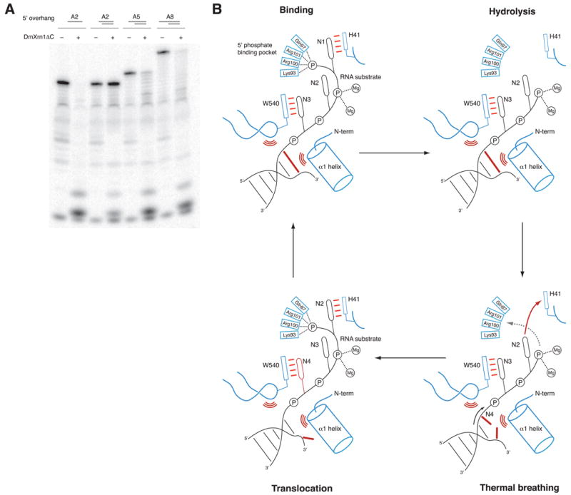
Messenger RNA decay plays a central role in the regulation and surveillance of eukaryotic gene expression. The conserved multidomain exoribonuclease Xrn1 targets cytoplasmic RNA substrates marked by a 5' monophosphate for processive 5'-to-3' degradation by an unknown mechanism. Here, we report the crystal structure of an Xrn1-substrate complex. The single-stranded substrate is held in place by stacking of the 5'-terminal trinucleotide between aromatic side chains while a highly basic pocket specifically recognizes the 5' phosphate. Mutations of residues involved in binding the 5'-terminal nucleotide impair Xrn1 processivity. The substrate recognition mechanism allows Xrn1 to couple processive hydrolysis to duplex melting in RNA substrates with sufficiently long single-stranded 5' overhangs. The Xrn1-substrate complex structure thus rationalizes the exclusive specificity of Xrn1 for 5'-monophosphorylated substrates, ensuring fidelity of mRNA turnover, and posits a model for translocation-coupled unwinding of structured RNA substrates.


Figures

Figure 1
Figure 1. Domain architecture of Drosophila melanogaster Xrn1 (DmXrn1)
(A) Schematic representation of DmXrn1 domains. The unstructured C-terminal region of DmXrn1 that was excluded from the crystallized construct is depicted in grey. (B) Overall view of DmXrn1ΔC-substrate complex, shown as ribbon (left) and surface (right) representations, with domains colored according to the schematic diagram in A. The nucleic acid substrate is colored black and shown in stick format. A Mg2+ ion is depicted as a purple sphere. Dashed lines indicate loops not resolved in electron density maps. Illustrations were generated using Pymol (Delano Scientific).
Figure 2
Figure 2. Xrn1 binds the N-terminal trinucleotide of the nucleic acid substrate
(A) The catalytic domain of DmXrn1 is shown in blue. The nucleic acid substrate is shown in black. The electron density map corresponds to the experimental SAD-phased map obtained after solvent flipping and phase extension to 3.2 Å, and is contoured at 1.0 σ. (B) Close-up view of substrate binding. The oligodeoxythymidine substrate is shown in stick format with carbon atoms colored white. The nucleotides are labeled N1–N3 in the 5′-3′ direction. Strictly conserved residues contacting the substrate are shown in stick format. (C) Surface representation of the active site of Xrn1, colored according to electrostatic potential. (D) A 5′-end-labeled G(CU)10C RNA oligonucleotide (200 nM) was pre-incubated in the presence or absence of DmXrn1ΔC (740 nM) and subsequently digested with RNase T1 and RNase A. Products were resolved by electrophoresis on a 16% polyacrylamide and 8 M urea gel, and visualized by phosphorimaging. The Xrn1-protected fragment is indicated with an arrow.
Figure 3
Figure 3. A conserved basic pocket in Xrn1 specifically recognizes the 5′ phosphate moiety of the substrate
(A) Close-up view of the 5′-phosphate-binding pocket. Strictly conserved residues are shown in stick format. (B) Single-turnover exonuclease activity assay of wild-type and mutant DmXrn1ΔC proteins. DmXrn1ΔC constructs (370 nM) were incubated with a 3′-[32P]-pCp labeled, 5′-phosphorylated G(CU)10C RNA oligonucleotide (200 nM) for 2 min at 20°C. Reactions were analyzed on a 16% denaturing gel, followed by phosphorimaging. (C) Wild-type (WT) and R100A/R101A DmXrn1ΔC proteins were incubated with 3′ end-labeled G(CU)10C oligonucleotides bearing either 5′-terminal phosphate or hydroxyl groups. Note that 5′-phosphorylation of the 3′-end-labeled substrate by polynucleotide kinase removes the 3′ phosphate group, resulting in different terminal products (CMP vs pCp). RNase T1 served as positive control for removal of a single nucleotide from the 5′ end. The slightly lower mobility of the RNase T1 digested fragment is due to the absence of a 5′ phosphate. (D) RNase protection assay of WT and R100A/R101A DmXrn1ΔC. The assay was performed as in Figure 2D.
Figure 4
Figure 4. Xrn1 couples hydrolysis to duplex unwinding in substrates with sufficiently long single-stranded 5′ extensions
(A) Hydrolysis-coupled duplex unwinding by DmXrn1ΔC. The substrates contain a common 17 base-pair GC-rich RNA/DNA duplex and a 2-, 5- or 8-nt oligo(A) overhang at the 5′ end of the RNA strand. The RNA strands were 3′-end labeled and 5′-phosphorylated. The two leftmost lanes represent control degradation of the A2 overhang RNA oligonucleotide in the absence of the complementary DNA strand. (B) Model of hydrolysis-coupled duplex unwinding by Xrn1. Following cleavage and release of the nucleotide monophosphate product, Xrn1 remains bound to substrate. Transient opening of the terminal base pair of the duplex by thermal breathing allows the substrate to advance through the narrow channel between the N-terminal helix α1 and the Trp540-containing loop. Substrate translocation occurs by a Brownian ratchet mechanism in which interactions of the 5′-terminal nucleotide with His41 and the 5′ phosphate binding pocket and subsequent hydrolysis impart directionality.

References

    1. Adams PD, Afonine PV, Bunkoczi G, Chen VB, Davis IW, Echols N, Headd JJ, Hung LW, Kapral GJ, Grosse-Kunstleve RW, et al. PHENIX: a comprehensive Python-based system for macromolecular structure solution. Acta Crystallogr D Biol Crystallogr. 2010;66:213–221. - PMC - PubMed
    1. Bonneau F, Basquin J, Ebert J, Lorentzen E, Conti E. The yeast exosome functions as a macromolecular cage to channel RNA substrates for degradation. Cell. 2009;139:547–559. - PubMed
    1. Chernyakov I, Whipple JM, Kotelawala L, Grayhack EJ, Phizicky EM. Degradation of several hypomodified mature tRNA species in Saccharomyces cerevisiae is mediated by Met22 and the 5′-3′ exonucleases Rat1 and Xrn1. Genes Dev. 2008;22:1369–1380. - PMC - PubMed
    1. Cole SE, LaRiviere FJ, Merrikh CN, Moore MJ. A convergence of rRNA and mRNA quality control pathways revealed by mechanistic analysis of nonfunctional rRNA decay. Mol Cell. 2009;34:440–450. - PMC - PubMed
    1. Coller J, Parker R. Eukaryotic mRNA decapping. Annu Rev Biochem. 2004;73:861–890. - PubMed

Publication types

Associated data