. 2011 Mar 4;8(3):309-17.
doi: 10.1016/j.stem.2011.01.012.

Short-term immunosuppression promotes engraftment of embryonic and induced pluripotent stem cells

Affiliations

Short-term immunosuppression promotes engraftment of embryonic and induced pluripotent stem cells

Jeremy I Pearl et al. Cell Stem Cell. .

Abstract

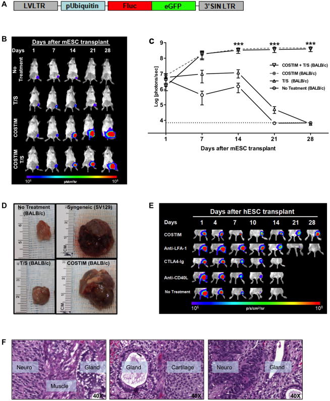
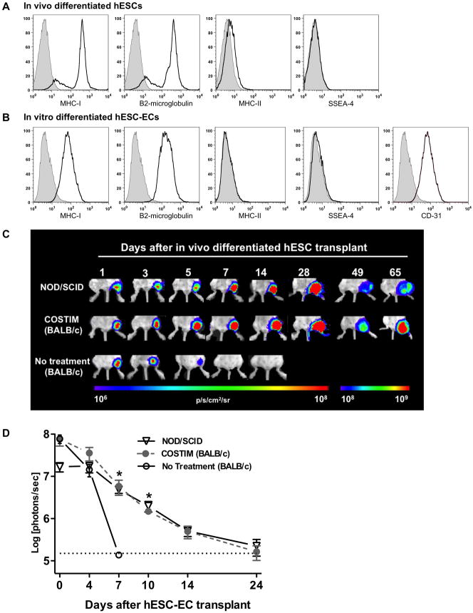
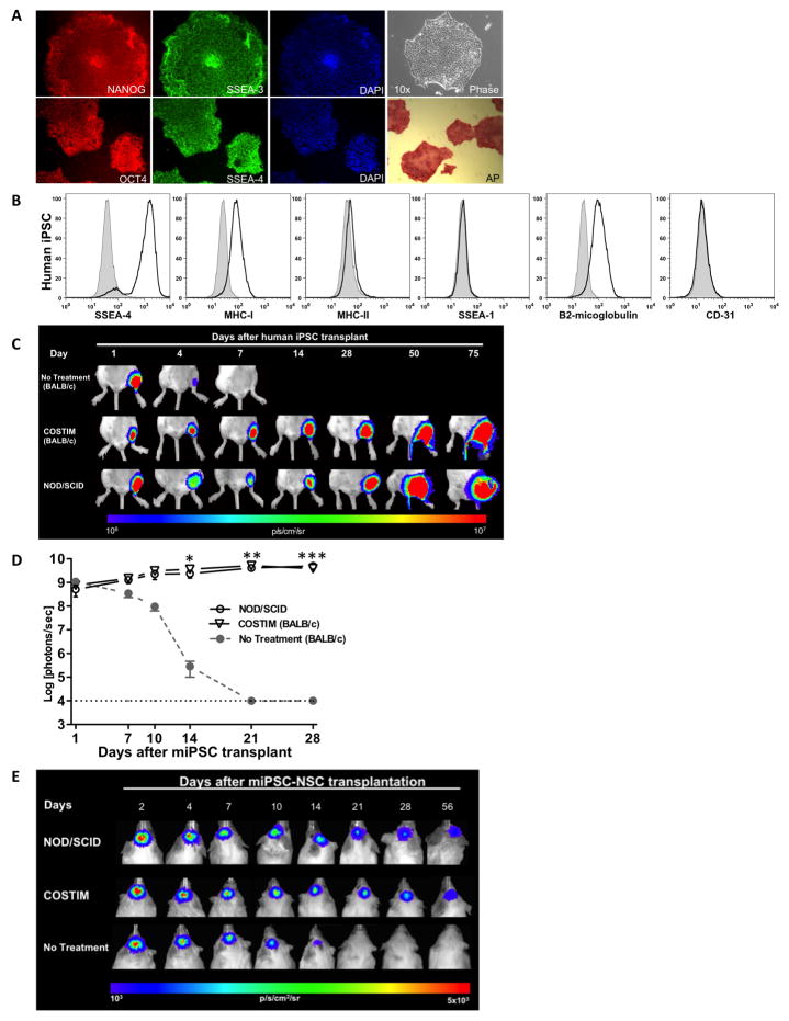
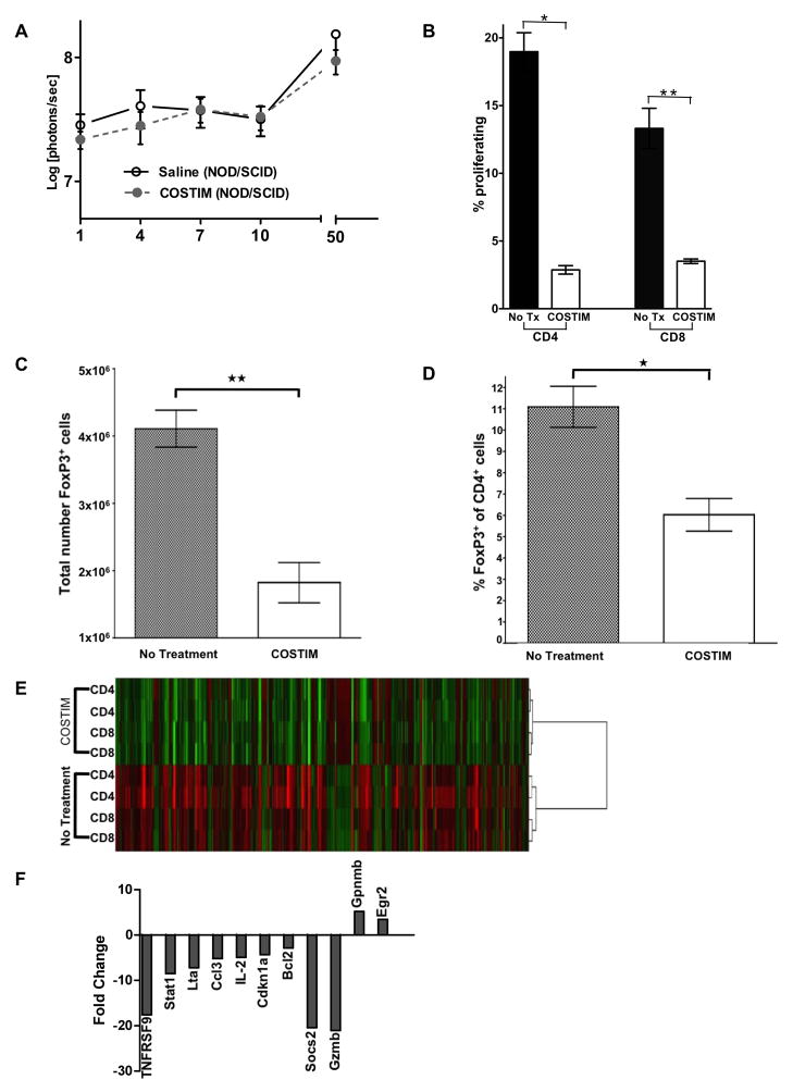
Embryonic stem cells (ESCs) are an attractive source for tissue regeneration and repair therapies because they can be differentiated into virtually any cell type in the adult body. However, for this approach to succeed, the transplanted ESCs must survive long enough to generate a therapeutic benefit. A major obstacle facing the engraftment of ESCs is transplant rejection by the immune system. Here we show that blocking leukocyte costimulatory molecules permits ESC engraftment. We demonstrate the success of this immunosuppressive therapy for mouse ESCs, human ESCs, mouse induced pluripotent stem cells (iPSCs), human induced pluripotent stem cells, and more differentiated ESC/(iPSCs) derivatives. Additionally, we provide evidence describing the mechanism by which inhibition of costimulatory molecules suppresses T cell activation. This report describes a short-term immunosuppressive approach capable of inducing engraftment of transplanted ESCs and iPSCs, providing a significant improvement in our mechanistic understanding of the critical role costimulatory molecules play in leukocyte activation.


Figures

Figure 1
Figure 1. Blockade of leukocyte costimulatory molecules mitigates allogeneic and xenogeneic transplantation rejection of undifferentiated ESCs
(a) Schema of the DF reporter gene construct containing Fluc and eGFP driven by a constitutive human ubiquitin promoter, using a self-inactivating (SIN) lentiviral vector. (b) Representative bioluminescence images and (c) quantitative bioluminescence intensity of mESC-transplanted mice that received either no treatment, tacrolimus + sirolimus (T/S), CTLA4-Ig + anti-LFA-1 + anti-CD40L (COSTIM), or COSTIM + T/S. n = 5 per group, ***P<0.001. (d) Representative images of gastrocnemius muscles 28 days after transplantation of non-transduced mESCs. (e) Representative bioluminescence images of xenogeneic hESC-transplanted mice that received no treatment, monotherapy, or a combination of all three costimulatory blockade agents (COSTIM). n = 5–8 per group. (f) Histopathological evaluation of HE-stained muscle sections from COSTIM treated mice demonstrating hESC-derived teratoma formation. All values are expressed as mean ± SEM. Color scale bars are in photons per second per centimeter squared per steradian (p/s/cm2/sr). H&E, hematoxylin and eosin stain. For further characterization of the ESCs and iPSCs, see Figure S1.
Figure 2
Figure 2. Leukocyte costimulatory molecule blockade permits engraftment of differentiated hESC-derivatives
Mean fluorescence intensity of MHC antigens, pluripotency (SSEA-4), and endothelial (CD31) markers on (a) in vivo differentiated hESCs isolated from explanted teratoma and (b) in vitro differentiated hESC-ECs. Filled histograms represent isotype control antibodies. (c) BLI of the survival of in vivo differentiated hESCs transplanted into immunodeficient (NOD/SCID) and immunocompetent (BALB/) mice that received either costimulatory blockade (COSTIM) or no immunosuppressive treatment, n = 3–4 per group. (d) Bioluminescence photon intensities representing the survival of in vitro differentiated hESC-ECs after transplantation into immunodeficient, costimulatory blockade (COSTIM) treated, or non-treated immunocompetent (BALB/c) mice, n = 4 per group, *P<0.05. For additional engraftment data regarding differentiated ESCs, see Figure S2.
Figure 3
Figure 3. Leukocyte costimulatory molecule blockade permits xenogeneic and allogeneic engraftment of hiPSC, miPSC, and differentiated miPSC-derivatives
(a) Characterization of hiPSCs by immunostaining with pluripotency markers such as Nanog, Oct4, SSEA-3, SSEA-4, and alkaline phosphatase (AP). (b) Mean fluorescence intensity of MHC antigens and pluripotency markers on undifferentiated hiPSCs. Filled histograms represent isotype control antibodies. BLI and bioluminescence photon intensities representing the survival of (c) hiPSCs and (d) miPSCs transplanted into the gastrocnemius muscle of immunodeficient (NOD/SCID) and immunocompetent mice receiving costimulatory blockade (COSTIM) or no treatment, n = 3–5 per group, *P<0.05, **P<0.01, ***P<0.001. (e) In vitro differentiated miPSC-NSCs transplanted into the subcortical area of the brain in immunodeficient (NOD/SCID) and immunocompetent mice. n = 3–4 per group. For additional characterization and engraftment data regarding miPSCs and hiPSCs, see Figure S3 and Movie S1.
Figure 4
Figure 4. Gene expression and functional characterization of leukocytes treated with costimulatory molecule blockade
(a) Bioluminescence photon intensities representing the survival of hESCs in immunodeficient (NOD/SCID) mice treated with COSTIM or saline as control. n = 5 per group. (b) Mixed lymphocyte reaction comparing the proliferation of COSTIM-treated and untreated T-cell subsets stimulated by allogeneic splenocytes. *P<0.0001, **P=0.0002. Shown is a representative trial chosen from three independent trials demonstrating similar results. (c) Comparison of the total number of CD4+FoxP3+ T cells and (d) percent of CD4+ cells that are FoxP3+ isolated from mice treated with COSTIM or saline as control. n = 6 COSTIM, n = 3 untreated control, *P=0.006, **P=0.002. (e) Hierarchical clustering of T-cells stimulated by allogeneic splenocytes reveals distinct gene expression clusters between COSTIM-treated and untreated T cells. Biological duplicates for each group are shown. (f) Gene expression fold change of COSTIM-treated relative to untreated T-cells. For additional characterization of the costimulatory blockade treated responder T-cells see Figure S4.

Comment in

References

    1. Abdullah Z, Saric T, Kashkar H, Baschuk N, Yazdanpanah B, Fleischmann BK, Hescheler J, Kronke M, Utermohlen O. Serpin-6 expression protects embryonic stem cells from lysis by antigen-specific CTL. J Immunol. 2007;178:3390–3399. - PubMed
    1. Assmus B, Honold J, Schachinger V, Britten MB, Fischer-Rasokat U, Lehmann R, Teupe C, Pistorius K, Martin H, Abolmaali ND, et al. Transcoronary transplantation of progenitor cells after myocardial infarction. N Engl J Med. 2006;355:1222–1232. - PubMed
    1. Bonde S, Zavazava N. Immunogenicity and engraftment of mouse embryonic stem cells in allogeneic recipients. Stem Cells. 2006;24:2192–2201. - PubMed
    1. Byrne JA. Generation of isogenic pluripotent stem cells. Hum Mol Genet. 2008;17:R37–41. - PubMed
    1. Cao F, Wagner RA, Wilson KD, Xie X, Fu JD, Drukker M, Lee A, Li RA, Gambhir SS, Weissman IL, et al. Transcriptional and functional profiling of human embryonic stem cell-derived cardiomyocytes. PLoS One. 2008;3:e3474. - PMC - PubMed

Publication types

MeSH terms

Associated data