. 2011 Apr 22;286(16):14575-87.
doi: 10.1074/jbc.M111.218990. Epub 2011 Mar 1.

Structure and biochemical functions of SIRT6

Affiliations

Structure and biochemical functions of SIRT6

Patricia W Pan et al. J Biol Chem. .

Abstract

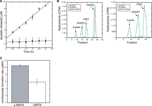
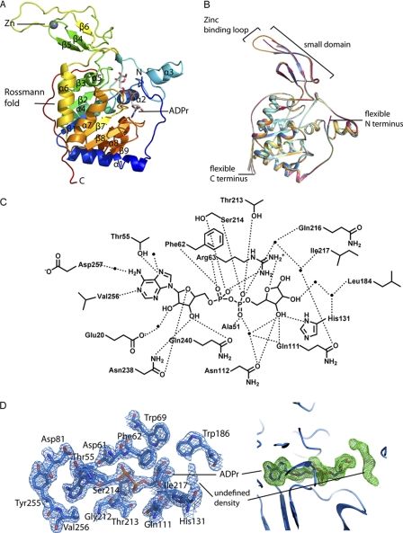
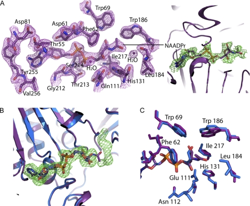
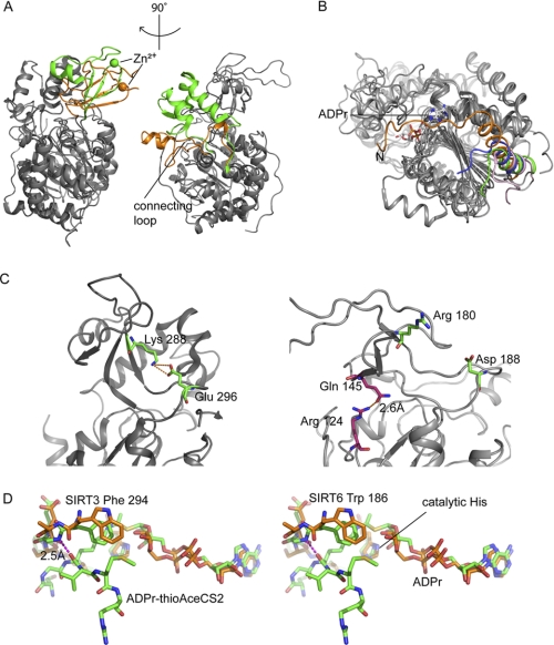
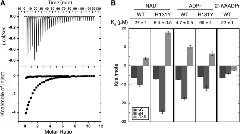
SIRT6 is a member of the evolutionarily conserved sirtuin family of NAD(+)-dependent protein deacetylases and functions in genomic stability and transcriptional control of glucose metabolism. Early reports suggested that SIRT6 performs ADP-ribosylation, whereas more recent studies have suggested that SIRT6 functions mainly as a histone deacetylase. Thus, the molecular functions of SIRT6 remain uncertain. Here, we perform biochemical, kinetic, and structural studies to provide new mechanistic insight into the functions of SIRT6. Utilizing three different assays, we provide biochemical and kinetic evidence that SIRT6-dependent histone deacetylation produces O-acetyl-ADP-ribose but at a rate ∼1,000 times slower than other highly active sirtuins. To understand the molecular basis for such low deacetylase activity, we solved the first crystal structures of this class IV sirtuin in complex with ADP-ribose and the non-hydrolyzable analog of O-acetyl-ADP-ribose, 2'-N-acetyl-ADP-ribose. The structures revealed unique features of human SIRT6, including a splayed zinc-binding domain and the absence of a helix bundle that in other sirtuin structures connects the zinc-binding motif and Rossmann fold domain. SIRT6 also lacks the conserved, highly flexible, NAD(+)-binding loop and instead contains a stable single helix. These differences led us to hypothesize that SIRT6, unlike all other studied sirtuins, would be able to bind NAD(+) in the absence of an acetylated substrate. Indeed, we found that SIRT6 binds NAD(+) with relatively high affinity (K(d) = 27 ± 1 μM) in the absence of an acetylated substrate. Isothermal titration calorimetry and tryptophan fluorescence binding assays suggested that ADP-ribose and NAD(+) induce different structural perturbations and that NADH does not bind to SIRT6. Collectively, these new insights imply a unique activating mechanism and/or the possibility that SIRT6 could act as an NAD(+) metabolite sensor.


Figures

FIGURE 1.
FIGURE 1.
SIRT6 deacetylation assays. A, charcoal-binding assay measuring the production of OAADPr. All assays were carried out in the presence of 4 μm SIRT6 WT (●) or H131Y (■), 300 μm [3H]H3K9Ac peptide, and 600 μm NAD+. Shown is the average of three experiments for WT and H131Y. The rate of OAADPr production was 3.2 ± 0.2 μm/h based on linear regression analysis with an R2 of 0.98612. B, HPLC separation of 3H-labeled products monitoring deacetylation of a [3H]H3K9Ac peptide. Reactions were carried out in the presence of 300 μm [3H]H3K9Ac peptide, 2 mm NAD+, and either 4 μm SIRT6, 4 μm Hst2 or no enzyme. Shown are all counts for the Hst2 deacetylation reaction (green), SIRT6 (brown), and a no enzyme control (blue) as well as a zoomed in view displaying counts for [3H]acetate, [3H]OAADPr, and H3K9 ([3H]acetylated N terminus). C, continuous assay monitoring the release of nicotinamide over time. Shown is the average of three trials with assays carried out in the presence of 8 μm (gray) or 0 μm (white) SIRT6, 300 μm H3K9Ac, and 0.6 mm NAD+. The rate of nicotinamide release in the presence of SIRT6 was 10.5 ± 0.4 μm/h and the rate in its absence was 5.9 ± 0.8 μm/h. Error bars, S.D.
FIGURE 2.
FIGURE 2.
Structure of human SIRT6 in complex with ADP-ribose. A, overall structural features of SIRT6 monomer. B, superimposition of the six molecules in the asymmetric unit. Red, chain A; green, chain B; dark blue, chain C; orange, chain D; cyan, chain E; yellow, chain F. C, schematic illustration of the hydrogen bonding network surrounding ADPr; hydrogen bonds are indicated as dashed lines, and water molecules are shown as spheres. D, left, SIRT6·ADPr 2FoFc electron density map (blue mesh, 1.5σ) of the residues within 4 Å of ADPr. Right, FoFc omit electron density map (green mesh, 2σ) of the ADPr molecule; the putative peptide binding site contains an unidentified electron density.
FIGURE 3.
FIGURE 3.
Comparison of SIRT6·ADP-ribose with SIRT6·2′-N-acetyl-ADP-ribose structure. A, left, SIRT6·NAADPr 2FoFc electron density map (purple mesh, 1.5σ) of the residues within 4 Å of NAADPr. Right, FoFc omit electron density map (green mesh, 1σ) of the NAADPr molecule. B, superimposition of SIRT6·ADPr (blue) onto SIRT6·NAADPr (purple); the unknown density (FoFc omit electron density map contoured at 2σ, green mesh) observed in the SIRT6·ADPr structure is not present in the SIRT6·NAADPr structure, and the N-acetyl group occupies part of the unidentified density space. C, residues immediately surrounding the N-acetyl group of NAADPr (purple) and their corresponding residues (blue) in the SIRT6·ADPr structure.
FIGURE 4.
FIGURE 4.
SIRT6·ADPr structure compared with other solved human SIRTs. A, SIRT6 structure (PDB code 3PKI, orange) superimposed onto SIRT3 (PDB code 3GLR, green). The left panel highlights differences in the zinc-binding domain, and the right panel shows a missing helix bundle. B, comparison of the N-terminal loop of SIRT6 (3PKI, orange), SIRT2 (1J8F, pink), SIRT3 (3GLR, green), and SIRT5 (2B4Y, blue); SIRT6 contains a long loop covering the NAD+ binding site. C, SIRT6 lacks the conserved salt bridge. Left, SIRT3 (3GLR) structure with conventional salt bridge in the sirtuin family. Right, the green pair (Arg180 and Asp188) is where the bridge would be predicted to form on the SIRT6 structure according to other sirtuin crystal structures. Pink pair (Arg124 and Gln145), the actual hydrogen bonding pair found in SIRT6 structure. D, stereo diagram of flipped FGEXL (WEDsL) loop of SIRT6 (PDB code 3PKI, orange) complexed with ADPr (orange) compared with SIRT3 (PDB code 3GLT, green) with an ADPr·thioAceCS2 (green) complex.
FIGURE 5.
FIGURE 5.
Isothermal titration calorimetry studies. A, representative ITC trace. The top graph shows data obtained for 37 automatic injections (1–8 μl) of 1.4 mm NAD+ titrated into 27 μm SIRT6. The bottom graph represents integrated curves of the experimentally generated heats. The data were fitted to a one-site binding curve (solid line). NAD+ (1.40 or 1.63 mm) was titrated into 27 μm WT SIRT6 (■) or 50 μm Hst2 (●), respectively. B, dissociation constants and column graph displaying thermodynamic parameters for WT and H131Y SIRT6 binding to NAD+, ADPr, and 2′-NAADPr. NAD+ (1.40 mm or 767 μm) was titrated into 27 μm or 33 μm WT or H131Y SIRT6, respectively. ADPr (390 μm or 1.45 mm) was titrated into 28 or 37 μm WT or H131Y SIRT6, respectively. 2′-NAADPr (2.96 mm) was titrated into 38 μm WT SIRT6. The change in Gibbs free energy (ΔG) is shown in black. Change in enthalpy (ΔH) is shown is gray, and −TΔS is displayed in light gray. Error bars, S.D.

References

    1. Michan S., Sinclair D. (2007) Biochem. J. 404, 1–13 - PMC - PubMed
    1. Jackson M. D., Denu J. M. (2002) J. Biol. Chem. 277, 18535–18544 - PubMed
    1. Sauve A. A., Celic I., Avalos J., Deng H., Boeke J. D., Schramm V. L. (2001) Biochemistry 40, 15456–15463 - PubMed
    1. Haigis M. C., Guarente L. P. (2006) Genes Dev. 20, 2913–2921 - PubMed
    1. Kaeberlein M., McVey M., Guarente L. (1999) Genes Dev. 13, 2570–2580 - PMC - PubMed

Publication types

LinkOut - more resources