Review
. 2011 Apr;17(2):197-208.
doi: 10.1177/1073858410386727. Epub 2011 Feb 28.

Laryngeal motor cortex and control of speech in humans

Affiliations
Review

Laryngeal motor cortex and control of speech in humans

Kristina Simonyan et al. Neuroscientist. 2011 Apr.

Abstract

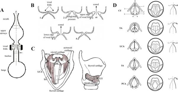
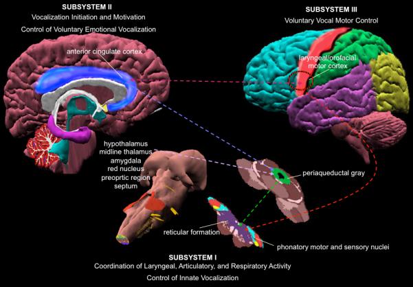
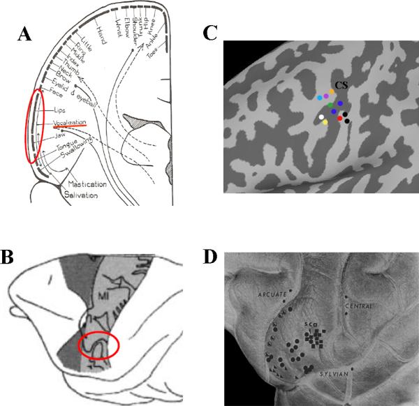
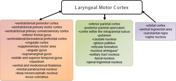
Speech production is one of the most complex and rapid motor behaviors, and it involves a precise coordination of more than 100 laryngeal, orofacial, and respiratory muscles. Yet we lack a complete understanding of laryngeal motor cortical control during production of speech and other voluntary laryngeal behaviors. In recent years, a number of studies have confirmed the laryngeal motor cortical representation in humans and have provided some information about its interactions with other cortical and subcortical regions that are principally involved in vocal motor control of speech production. In this review, the authors discuss the organization of the peripheral and central laryngeal control based on neuroimaging and electrical stimulation studies in humans and neuroanatomical tracing studies in nonhuman primates. It is hypothesized that the location of the laryngeal motor cortex in the primary motor cortex and its direct connections with the brain stem laryngeal motoneurons in humans, as opposed to its location in the premotor cortex with only indirect connections to the laryngeal motoneurons in nonhuman primates, may represent one of the major evolutionary developments in humans toward the ability to speak and vocalize voluntarily.


Figures

Figure 1
Figure 1. (A) Schematic view of the vocal and respiratory tracts
Voice originates in the larynx. First, the expiratory airflow from the lungs reaches the larynx through the trachea, where it sets the closed vocal fold tissue into self-excited oscillatations, due to which the larynx becomes the source of voice sound. Further, pressure from the vocal fold oscillations is resonated through the vocal tract and radiated from the mouth as voice. (B) Schematic sequence of events preceding voice production. (1). The vocal folds close immideatly prior to voice production; (2) subglottal air pressure builds up below the vocal folds during exhalation; (3) lower and upper edge of the vocal folds separate subsequently with the release of air and sound generation; (4) the vocal folds re-approximate, starting from their lower edge, and (5) the vocal folds close completely before the next sound production. (C) Superior and lateral views of the human larynx. Intrinsic laryngeal muscles and cartilages. TA – thyroarytenoid muscle; LCA – lateral cricoarytenoid muscle; PCA – posterior cricoarytenoid muscle; IA – interarytenoid muscle; CT - cricothyroid muscle. The arrows show the directions of the muscle contractions. (D) Schematic presentation of the laryngeal muscle function. The left column shows the location of the cartilages and the edge of the vocal folds when each of the laryngeal muscles is active. The arrows indicate the directions of the force exerted. 1. thyroid cartilage; 2. cricoid cartilage; 3. arytenoid cartilages; 4. vocal ligament; 5. posterior cricoarytenoid ligament. The middle column shows the laryngeal view. The right column shows contours of the frontal section at the middle of the membranous portion of the vocal fold. The dotted line shows a state in which no muscle is activated (reprinted from Hirano, 1981 with permission of Springer Science + Business Media).
Figure 2
Figure 2. Hierarchical organization of central voice control in humans and non-human primates
The figure depicts different levels of the voice control system. The lowest level (Subsystem I) is represented by the brainstem and spinal cord sensorimotor phonatory nuclei. This subsystem is responsible for the coordination of laryngeal, articulatory and respiratory control during production of innate vocalizations. The higher level within this system (Subsystem II) is represented by the PAG, ACC and limbic input structures, such as the hypothalamus, midline thalamus, amygdala, red nucleus, preoptic region, septum. This subsystem is responsible for initiation of vocalizations and control of voluntary emotional vocalizations. The highest level is represented by the laryngeal/orofacial motor cortex with its input and output regions (Subsystem III). This subsystem is responsible for voluntary vocal motor control of speech and song production. The dotted lines show simplified connections between different regions within the voice controlling system.
Figure 3
Figure 3. Laryngeal motor cortical representation
Schematic views of body representation within the motor cortex (A) in humans (“motor homunculus” according to (Penfield and Bordley, 1937)) and (B) in the rhesus monkey (“motor simiculus” according to (Woolsey et al., 1952), reprinted from Fadiga et al., 2000 with permission from Elsevier). (C) The laryngeal motor cortical region in humans as defined in neuroimaging studies. The colored circles represent the reported peaks of activation in the following studies of syllable production: orange - (Bohland and Guenther, 2006); purple - (Olthoff et al., 2008); light blue - (Terumitsu et al., 2006); green - (Loucks et al., 2007); blue - (Brown et al., 2008); red -(Wilson et al., 2004); black - (Simonyan et al., 2009); white - (Riecker et al., 2008), and yellow - (Peeva et al.). CS - central sulcus. (D) The laryngeal motor cortical region in the rhesus monkey. Topographical representation of the laryngeal muscles: cricothyroid - right-angled triangle; thyroarytenoid - circle; combination of the circothyroid and thyroarytenoid - encircled right-angled triangle, and extrinsic laryngeal muscles - square. sca - sulcus subcentralis anterior (subcentral dimple) (reprinted from (Hast et al., 1974) with permission from Elsevier).
Figure 4
Figure 4. Cortical and subcortical networks of the laryngeal motor cortex
Block diagrams illustrate the reciprocal (pink box), outgoing (green box) and incoming (purple box) connections of the laryngeal motor cortex as defined using neuroanatomical tracing studies in non-human primates and diffusion tensor tractography in humans. Asterisk (*) indicates that projection from the laryngeal motor cortex to the nucleus ambiguus exists only in humans but not in non-human primates.

References

    1. Alario FX, Chainay H, Lehericy S, Cohen L. The role of the supplementary motor area (SMA) in word production. Brain Res. 2006;1076:129–143. - PubMed
    1. Bauer G, Gerstenbrand F, Hengl W. Involuntary motor phenomena in the locked-in syndrome. J Neurol. 1980;223:191–198. - PubMed
    1. Bernard JF, Villanueva L, Carroue J, Le Bars D. Efferent projections from the subnucleus reticularis dorsalis (SRD): a Phaseolus vulgaris leucoagglutinin study in the rat. Neurosci Lett. 1990;116:257–262. - PubMed
    1. Bohland JW, Guenther FH. An fMRI investigation of syllable sequence production. Neuroimage. 2006;32:821–841. - PubMed
    1. Brown S, Laird AR, Pfordresher PQ, Thelen SM, Turkeltaub P, Liotti M. The somatotopy of speech: phonation and articulation in the human motor cortex. Brain Cogn. 2009;70:31–41. - PMC - PubMed

Publication types

LinkOut - more resources