. 2011 Mar 1;71(5):1584-96.
doi: 10.1158/0008-5472.CAN-10-3166.

Stromal deletion of the APC tumor suppressor in mice triggers development of endometrial cancer

Affiliations

Stromal deletion of the APC tumor suppressor in mice triggers development of endometrial cancer

Pradeep S Tanwar et al. Cancer Res. .

Abstract

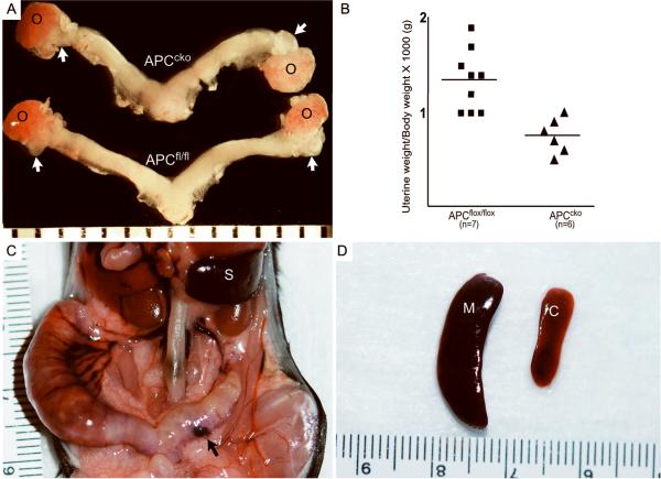
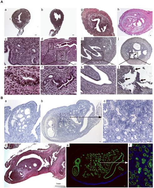
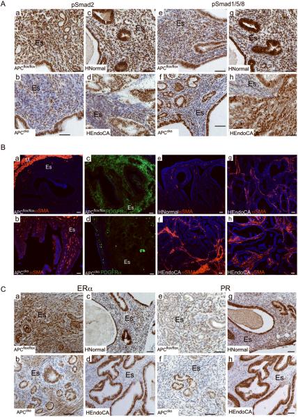
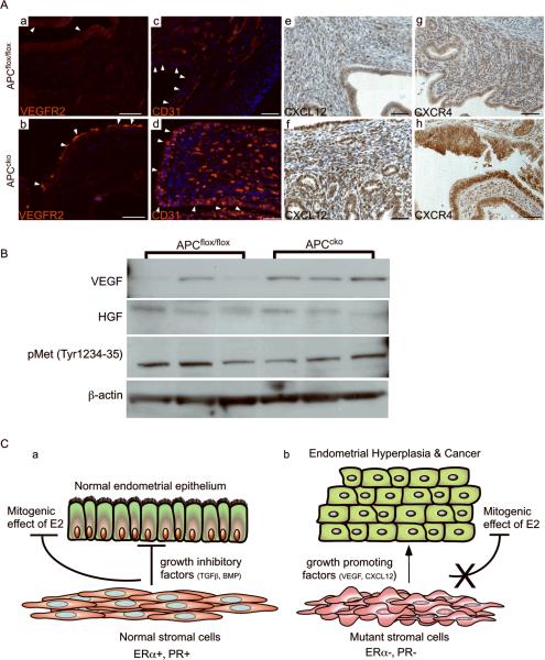
The contribution of the stromal microenvironment to the progression of endometrial cancer has not been well explored. We have conditionally expressed a mutant allele of adenomatous polyposis coli (APC(cKO)) in murine uterine stroma cells to study its effect on uterine development and function. In addition to metrorrhagia, the mice develop complex atypical endometrial gland hyperplasia that progresses to endometrial carcinoma in situ and endometrial adenocarcinoma as evidenced by myometrial invasion. Stromal cells subjacent to the carcinoma cells express alpha-smooth muscle actin (αSMA) with fewer cells expressing platelet-derived growth factor α compared with normal stromal cells, suggesting that the mutant stromal cells have acquired a more myofibroblastic phenotype, which have been described as cancer-associated fibroblasts and have been shown to induce carcinogenesis in other organ systems. Analyses of human endometrial cancer specimens showed substantial αSMA expression in the stroma compared with normal endometrial stroma cells. We also show that APC(cKO) mutant uteri and human endometrial cancer have decreased stromal levels of transforming growth factor β and bone morphogenetic protein activities and that the mutant uteri failed to respond to exogenous estradiol stimulation. The mutant stroma cells also had higher levels of vascular endothelial growth factor and stromal derived factor signaling components and diminished expression of estrogen receptor α and progesterone receptor, which is common in advanced stages of human endometrial cancer and is an indicator of poor prognosis. Our results indicate that de novo mutation or loss of heterozygosity in stromal APC is sufficient to induce endometrial hyperplasia and endometrial carcinogenesis by mechanisms that are consistent with unopposed estrogen signaling in the endometrial epithelium.


Figures

Figure 1
Figure 1
Analyses of Amhr2-Cre-induced recombination in murine uteri. (Panel A) Amhr2-Cre mice were crossed with LacZ and Yfp reporter mice. Amhr2-Cre induced β-galactosidase activity and direct YFP fluorescence was detected in stroma (ES) but not in epithelial cells of uteri (arrowheads and asterisks) or in their respective controls (A, insets). (Panel B, a) PCR of genomic DNA collected from the epithelium and stroma of APCcko tumors using laser capture microdissection and from whole uterus, ovary, oviduct, and tail was used to detect recombined 500 bp floxed allele. The unrecombined flox APC allele (430 bp) was present in all tissue examined. (Panel B, b) Western blot analyses of uterine lysates showing increased expression of β-catenin, TCF1, LEF1, and Cyclin d1 in APCcko compared to control mice. β-actin was used a loading control. (Panel C) Immunolocalization of β-catenin in 4-wk-old (a, b) and 5-month old (c, d) uteri of control and APCcko mice. Inset in b is a higher magnification image of area outlined by dotted lines in same panel. Arrowheads in panel d show nuclear accumulation of β-catenin in stroma. Epi: epithelium; ES: endometrial stroma. Bar equals 50 μm.
Figure 2
Figure 2
Gross phenotypic changes in the reproductive tracts of APCflox/flox, APCcko female mice. (Panel A) 4wk old uteri from representative APCflox/flox and APCcko mice are shown. White arrows point to the oviducts of control and mutant mice, which are shorter and exhibit less coiling in mutants. (Panel B) The average uterine wet weight of 4-week-old APCcko/ mice was also significantly smaller (p < 0.05) when compared with controls (0.75 g ± 0.76 vs. 1.34 g ± 0.11, respectively). (Panel C) Grossly enlarged APCcko uteri with blood filled cyst (black arrow) (D) Splenomegaly in APCcko mice compared to controls. o: ovary; M: mutant; C: control; S: spleen.
Figure 3
Figure 3
Histological analyses of uteri collected at different developmental stages from APCflox/flox and APCcko mice. Cross sections (Panel A, a & b) and longitudinal sections (Panel A, c & d) of 4wk old control (Panel A, a & c) and mutant (Panel A, b & d) uteri. Rectangular areas in Panel A, d are shown in higher magnification in e and f. Hyperplasia of epithelial lining of 5 month old APCcko mice uteri (Panel A, h) is shown compared with control (Panel A, g). (Panel A, i & k), polyp like outgrowths (Arrow) were observed from endometrium of some 7-month old mutant uteri (Panel A, j & l). Panel B, a shows development of carcinoma in situ in APCcko uteri. Panel B, b–d show mutant uteri with occlusion of uterine lumen by carcinogenic growth. Higher magnification image from Panel B, b showing admixed endometrial epithelial and stroma cells is shown in Panel B, c. Staining of mutant uterus with cytokeratin 8, an epithelial cell-specific marker. (Panel B, e). The boxed area is shown at higher magnification in Panel B, f. Bar equals 50 μm unless otherwise indicated.
Figure 4
Figure 4
Myometrial invasion by endometrial adenocarcinoma in adult APCcko mice. CD133 expression, which is highly expressed in human endometrial carcinoma, was detected at higher levels in endometrial tumors of APCcko mice compared with controls (Panels A & B). Inset in Panel B is a higher magnification of area boxed in white dashed line. H&E sections of 8 month-old APCcko uteri with endometrial adenocarcinoma showing fully formed endometrial glands (arrows) present in the myometrium (M) (Panels C–D). (Panels E & F) Colocalization of αSMA (red, a smooth muscle specific marker) and Cytokeratin 8 (ck, green, an epithelial specific marker) confirmed the presence of endometrial glands (white arrows) in the myometrium. Control uteri are shown in Panels G & H. White dotted line in Panel C shows demarcation of smooth muscle layer from the endometrial stromal compartment. Bar equals 50 μm.
Figure 5
Figure 5
APCcko uterine stroma is insensitive to estradiol treatment. APCflox/flox uterine size (Panel A, a) and weight (Panel B) is greatly increased after treatment compared to APCcko (Panel A, b & B) mice uteri. Histological sections from untreated control (Panel A, c) and mutant (Panel A, d) uteri were compared with age-matched, estradiol-treated control (Panel A, e) and mutant (Panel A, f) uteri. (Panel C) Western blot analyses of ERα, PR, and Foxo1 in mutant and control uterine lysates (n=3 each). β-actin was used as a loading control. Bar equals 50 μm.
Figure 6
Figure 6
APC deletion suppresses paracrine signaling and increases the myofibroblast population in mutant uterine stroma. (Panel A) Reduced expression of pSmad2 (a–d) and pSmad 1/5/8 (e–h) in the stroma, but not the epithelium, of mutant uteri and in human EC when compared with control uteri and in normal human endometrium was obaserved by IHC. (Panel B) Immunofluorescence was performed for αSMA (a & b) and PDGFRα (c & d) on 7 month-old APCflox/flox and APCcko uterus. In normal/benign human endometrium (n=4), αSMA is normally detected in vascular smooth muscle cells only (e) but in patients with EC (7/9), αSMA was detected in stromal cells (f–h). (Panel C) Suppression of ERα (a–d) and PR (e–h) expression in stroma of APCcko uteri and human EC patients compared to control or normal tissues was observed by IHC. HNormal: normal human endometrium HEndoCA: human EC. ES: endometrial stroma; M: myometrium. Bar equals 50 μm.
Figure 7
Figure 7
Increased carcinogenic signaling in APCcko uteri. (Panel A, a &b) Expression of VEGFR2 on the leading edge of tumor (arrowheads) by immunofluorescence in APCcko mice compared to controls (arrowheads). Increased numbers of CD31 positive-endothelial cells were present in mutant uteri (arrowheads) compared to controls (arrowheads) by immunofluorescence (c & d). Increased expression of CXCL12 in stroma (e & f) and CXCR4 in epithelium (g & h) in APCcko mice by IHC compared with controls. (Panel B) Western blot analyses of VEGF, HGF, pMet (tyr 1234–1235) and β-actin in control and mutant APCcko uteri. (Panel C) Schematic summarizing the effect of stromal-specific APC deletion on epithelial cells. Normal fibroblastic stromal cells (PR+ and ERα+) maintain uterine epithelial homeostasis by secreting growth inhibitory factors (TGFβ and BMPs) to regulate the mitogenic effect of estrogen on epithelial proliferation (a). Mutant myofibroblastic stromal cells (PR-neg and ERα~neg) express reduced levels of growth inhibitory factors (TGFβ and BMPs) and express more growth-promoting factors (VEGF and CXCL12), and are unable to oppose estrogen-induced ERα+ epithelial proliferation, which leads to hyperplasia and carcinogenesis. Bar equals 50 μm.

References

    1. Bissell MJ, Radisky D. Putting tumours in context. Nat Rev Cancer. 2001;1:46–54. - PMC - PubMed
    1. Cunha GR, Hayward SW, Wang YZ. Role of stroma in carcinogenesis of the prostate. Differentiation. 2002;70:473–85. - PubMed
    1. Kurita T, Cooke PS, Cunha GR. Epithelial-stromal tissue interaction in paramesonephric (Mullerian) epithelial differentiation. Dev Biol. 2001;240:194–211. - PubMed
    1. Booth BW, Mack DL, Androutsellis-Theotokis A, McKay RD, Boulanger CA, Smith GH. The mammary microenvironment alters the differentiation repertoire of neural stem cells. Proc Natl Acad Sci U S A. 2008;105:14891–6. - PMC - PubMed
    1. Boulanger CA, Mack DL, Booth BW, Smith GH. Interaction with the mammary microenvironment redirects spermatogenic cell fate in vivo. Proc Natl Acad Sci U S A. 2007;104:3871–6. - PMC - PubMed

Publication types

MeSH terms