. 2011 May;152(5):1989-2000.
doi: 10.1210/en.2010-1396. Epub 2011 Mar 1.

Thyroid hormone regulates the expression of the sonic hedgehog signaling pathway in the embryonic and adult Mammalian brain

Affiliations

Thyroid hormone regulates the expression of the sonic hedgehog signaling pathway in the embryonic and adult Mammalian brain

Lynette A Desouza et al. Endocrinology. 2011 May.

Abstract

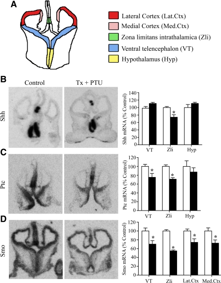
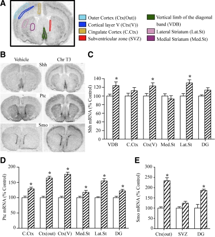
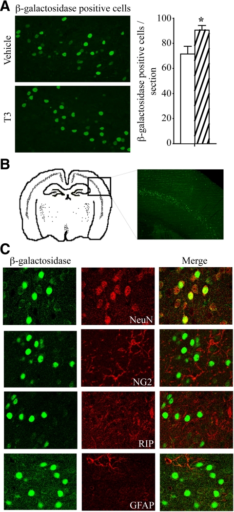
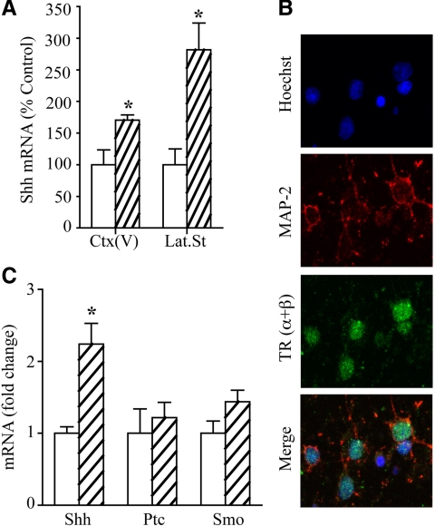
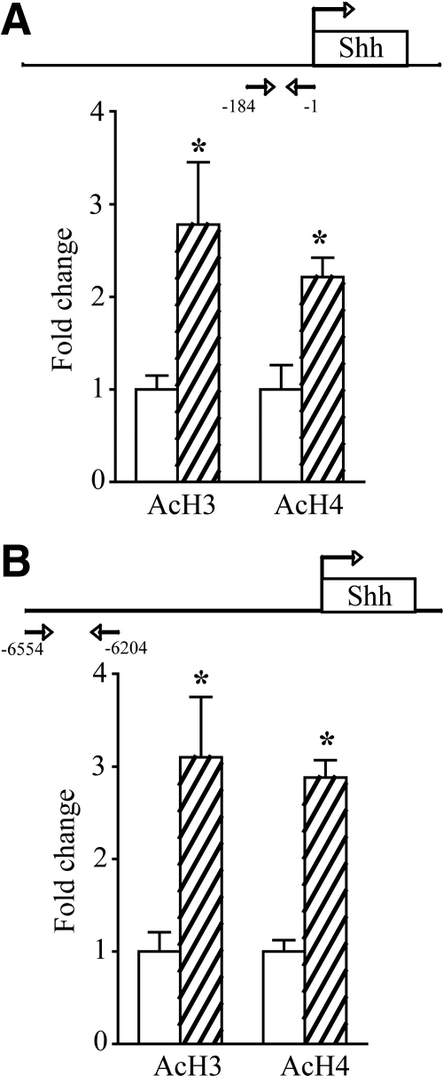
Thyroid hormone is important for development and plasticity in the immature and adult mammalian brain. Several thyroid hormone-responsive genes are regulated during specific developmental time windows, with relatively few influenced across the lifespan. We provide novel evidence that thyroid hormone regulates expression of the key developmental morphogen sonic hedgehog (Shh), and its coreceptors patched (Ptc) and smoothened (Smo), in the early embryonic and adult forebrain. Maternal hypo- and hyperthyroidism bidirectionally influenced Shh mRNA in embryonic forebrain signaling centers at stages before fetal thyroid hormone synthesis. Further, Smo and Ptc expression were significantly decreased in the forebrain of embryos derived from hypothyroid dams. Adult-onset thyroid hormone perturbations also regulated expression of the Shh pathway bidirectionally, with a significant induction of Shh, Ptc, and Smo after hyperthyroidism and a decline in Smo expression in the hypothyroid brain. Short-term T₃ administration resulted in a significant induction of cortical Shh mRNA expression and also enhanced reporter gene expression in Shh(+/LacZ) mice. Further, acute T₃ treatment of cortical neuronal cultures resulted in a rapid and significant increase in Shh mRNA, suggesting direct effects. Chromatin immunoprecipitation assays performed on adult neocortex indicated enhanced histone acetylation at the Shh promoter after acute T₃ administration, providing further support that Shh is a thyroid hormone-responsive gene. Our results indicate that maternal and adult-onset perturbations of euthyroid status cause robust and region-specific changes in the Shh pathway in the embryonic and adult forebrain, implicating Shh as a possible mechanistic link for specific neurodevelopmental effects of thyroid hormone.


Figures

Fig. 1.
Fig. 1.
Maternal hypothyroidism decreases Shh signaling cascade expression in the embryonic rat brain. Rat e13.5 pups were obtained from thyroidectomized females that were administered PTU (Tx + PTU), and levels of Shh, Ptc, and Smo mRNA were determined by in situ hybridization. Shown is a schematic representation of a coronal section through the embryonic rat brain with a color code depicting the regions quantitated (A) and representative autoradiographs of Shh (B), Ptc (C), and Smo (D) mRNA in embryos derived from control and hypothyroid (Tx + PTU) female dams. Maternal hypothyroidism resulted in a significant decrease in Shh mRNA in the Zli with no change in the VT or Hyp (B). Ptc and Smo mRNA were significantly down-regulated in several forebrain regions (namely the VT, Zli, Lat.Ctx and Med.Ctx) of e13.5 embryos derived from hypothyroid dams (C and D). Results are expressed as a percentage of control and are the mean ± sem (n = 11–15 embryos per group derived from four to five dams in each group). *, P < 0.05 compared with embryos derived from control dams (Student's unpaired t test). White bars, Embryos derived from vehicle-treated dams; black bars, embryos derived from Tx + PTU-treated dams.
Fig. 2.
Fig. 2.
Maternal hyperthyroidism enhances the expression of Shh in the embryonic rat brain. Rat e13.5 pups were derived from vehicle-treated and chronic T3-treated dams, and the levels of Shh, Ptc, and Smo mRNA were determined by in situ hybridization. Shown are representative autoradiographs showing Shh mRNA expression (A) in the VT, Zli, and Hyp in e13.5 embryos derived from vehicle or T3-treated dams. Maternal hyperthyroidism resulted in a significant induction in Shh mRNA in the VT, Zli, as well as Hyp, of the embryonic brain (A). Ptc and Smo mRNA remained unaltered in embryos derived from maternally hyperthyroid dams (B and C). Results are expressed as a percentage of control and are the mean ± sem (n = 7/group derived from three to four dams in each group). *, P < 0.05 when compared with control (Student's unpaired t test). White bars, embryos derived from vehicle-treated dams; black bars, embryos derived from chronic T3-treated dams.
Fig. 3.
Fig. 3.
Adult-onset hypothyroidism decreases Smo mRNA expression in the DG subfield of the hippocampus. Adult male rats were rendered hypothyroid by administration of MMI for 28 d, or PTU for 21 d, and the levels of Smo mRNA were determined by in situ hybridization. Shown are schematic representations of coronal sections through the ventricular zone and hippocampus of the adult rodent brain highlighting the SVZ and the DG region of the hippocampus, respectively (A). Smo mRNA was significantly and selectively down-regulated in the DG after adult-onset hypothyroidism compared with vehicle-treated controls (B). Results are expressed as a percentage of control and are the mean ± sem (n = 4/group). *, P < 0.05 compared with control (one-way ANOVA, Bonferroni post hoc test). White bars, Vehicle-treated controls; gray bars, MMI-treated animals; black bars, PTU-treated animals.
Fig. 4.
Fig. 4.
Shh mRNA is significantly up-regulated in the adult rat brain in response to chronic T3 administration. Adult male rats received chronic T3 (Chr T3) administration for a period of 10 d to render them hyperthyroid, and the levels of Shh, Ptc, and Smo mRNA were determined by in situ hybridization. Shown is a schematic representation of a coronal section of the adult rat brain with a color code depicting the regions quantitated (A). Also shown are representative autoradiographs of Shh, Ptc, and Smo mRNA expression in coronal sections of the adult brains from vehicle and chronic T3-treated male rats (B). Adult-onset hyperthyroidism resulted in a robust and significant increase in Shh expression in the VDB, cortex, and Lat.St (C). Chronic T3 treatment significantly increased the expression of Ptc (D) and Smo (E) mRNA in the adult rat brain. Results are expressed as a percentage of control and are the mean ± sem (n = 5–6/group). *, P < 0.05 compared with control (Student's unpaired t test). White bars, Vehicle-treated controls; hatched bars, chronic T3-treated animals.
Fig. 5.
Fig. 5.
Subchronic T3 treatment enhances the number of β-galactosidase immunopositive cells within the neocortex of Shh+/LacZ mice. Shown are representative images of β-galactosidase expressing cells in the cortex after short-duration T3 treatment over 2 d in Shh+/LacZ mice. T3 treatment significantly increased the number of cells that were strongly immunopositive for β-galactosidase in layer V of cortex (A). Results are expressed as a percentage of vehicle-treated control and are the mean ± sem (n = 5/group). *, P < 0.05 compared with control (Student's unpaired t test). Shown is a schematic representation (B) of a coronal section illustrating β-galactosidase immunopositive cells in Shh+/LacZ mice. The boxed area is enlarged to show a representative area indicating the presence of several strongly immunopositive β-galactosidase immunopositive cells in the neocortex of T3-treated Shh+/LacZ mice. Double immunofluorescence experiments revealed the colocalization of β-galactosidase with the mature neuronal marker NeuN but not with the astroglial (GFAP) or oligodendrocytic (RIP or NG2) markers examined (C).
Fig. 6.
Fig. 6.
Acute T3 treatment increases Shh mRNA expression in vivo and in vitro. Adult male rats received a single injection of T3, and the levels of Shh mRNA were determined 3 h later by in situ hybridization. Acute T3 treatment resulted in a significant increase in Shh mRNA expression in the cortex and Lat.St (A). Results are expressed as a percentage of control and are the mean ± sem (n = 3–5/group). *, P < 0.05 compared with vehicle-treated controls (Student's unpaired t test). Ctx(V), Cortical layer V. Shh, Ptc, and Smo mRNA was determined by qPCR from T3-treated primary cortical neuron cultures, isolated from e17.5 pups that were grown in vitro for 9 d before acute treatment with 20 nm T3 for 3 h. Shown are representative images of MAP-2 immunopositive primary cortical neurons expressing all TR isoforms (B) with their nuclei counterstained with Hoechst 33342. Acute T3 treatment of cortical cultures resulted in a significant increase in Shh mRNA expression, whereas Ptc and Smo mRNA remained unchanged as revealed by qPCR analysis (C). All genes were normalized to the housekeeping gene Hprt. Results are expressed as fold change and are the mean ± sem (n = 3–5/group). *, P < 0.05 compared with vehicle-treated controls (Student's unpaired t test).
Fig. 7.
Fig. 7.
Acute T3 treatment increases histone acetylation within upstream regulatory regions of the Shh gene in the adult rat cortex. ChIP assays were performed for pan histone H3 and H4 acetylation changes within gene regulatory sequences from −184 bp to the transcriptional start site of the Shh gene. Cortical tissue derived from T3-treated animals had significantly enhanced acetylation of histone H3 and H4 (AcH3 and AcH4) close to the transcriptional start site within the Shh promoter after acute T3 treatment (A). ChIP analysis within upstream regions of the Shh gene that contained putative TREs (−6554 to −6204 bp) revealed that cortical tissue derived from T3-treated animals had increased acetylation of both histone H3 and H4 within these regulatory sequences as well (B). Results are expressed as fold change and are the mean ± sem (n = 7–10/group). *, P < 0.05 compared with vehicle-treated controls (Student's unpaired t test).

Similar articles

Cited by

References

    1. Porterfield SP, Hendrich CE. 1993. The role of thyroid hormones in prenatal and neonatal neurological development—current perspectives. Endocr Rev 14:94–106 - PubMed
    1. Anderson GW, Schoonover CM, Jones SA. 2003. Control of thyroid hormone action in the developing rat brain. Thyroid 13:1039–1056 - PubMed
    1. Anderson GW. 2001. Thyroid hormones and the brain. Front Neuroendocrinol 22:1–17 - PubMed
    1. Akaike M, Kato N, Ohno H, Kobayashi T. 1991. Hyperactivity and spatial maze learning impairment of adult rats with temporary neonatal hypothyroidism. Neurotoxicol Teratol 13:317–322 - PubMed
    1. Williams GR. 2008. Neurodevelopmental and neurophysiological actions of thyroid hormone. J Neuroendocrinol 20:784–794 - PubMed

Publication types

MeSH terms