. 2011 Mar 1;25(5):445-59.
doi: 10.1101/gad.2014311.

Reduced fidelity of branch point recognition and alternative splicing induced by the anti-tumor drug spliceostatin A

Affiliations

Reduced fidelity of branch point recognition and alternative splicing induced by the anti-tumor drug spliceostatin A

Anna Corrionero et al. Genes Dev. .

Abstract

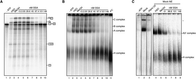
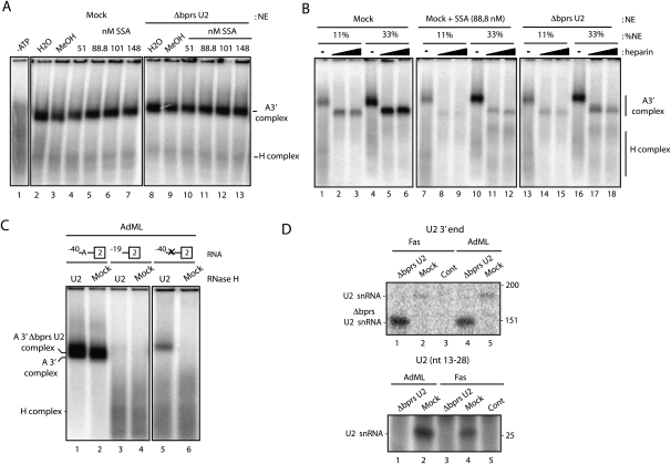
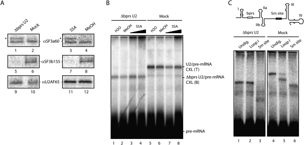
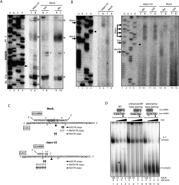
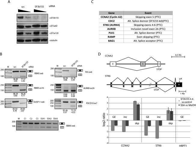
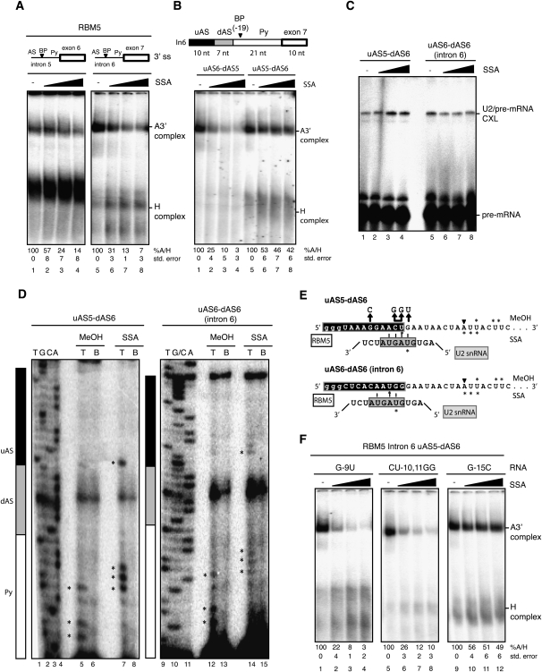
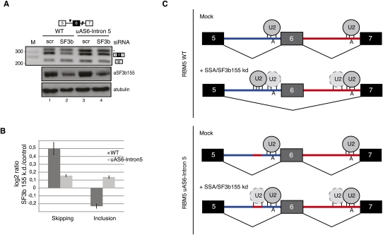
Spliceostatin A (SSA) is a stabilized derivative of a Pseudomonas bacterial fermentation product that displays potent anti-proliferative and anti-tumor activities in cancer cells and animal models. The drug inhibits pre-mRNA splicing in vitro and in vivo and binds SF3b, a protein subcomplex of U2 small nuclear ribonucleoprotein (snRNP), which is essential for recognition of the pre-mRNA branch point. We report that SSA prevents interaction of an SF3b 155-kDa subunit with the pre-mRNA, concomitant with nonproductive recruitment of U2 snRNP to sequences 5' of the branch point. Differences in base-pairing potential with U2 snRNA in this region lead to different sensitivity of 3' splice sites to SSA, and to SSA-induced changes in alternative splicing. Indeed, rather than general splicing inhibition, splicing-sensitive microarray analyses reveal specific alternative splicing changes induced by the drug that significantly overlap with those induced by knockdown of SF3b 155. These changes lead to down-regulation of genes important for cell division, including cyclin A2 and Aurora A kinase, thus providing an explanation for the anti-proliferative effects of SSA. Our results reveal a mechanism that prevents nonproductive base-pairing interactions in the spliceosome, and highlight the regulatory and cancer therapeutic potential of perturbing the fidelity of splice site recognition.


Figures

Figure 1
Figure 1
SSA inhibits U2 snRNP-containing A complex assembly. (A) In vitro splicing assays using AdML RNA in the presence of the indicated concentrations of SSA or its carrier (methanol), analyzed by denaturing polyacrylamide gel electrophoresis. Pre-mRNA, splicing intermediates, and products are indicated. (B) Spliceosome assembly assays corresponding to in vitro splicing mixes as in A in the presence of heparin (5 μg/μL), analyzed by native agarose–polyacrylamide gel electrophoresis. The positions of heterogeneous nuclear RNP (H) and spliceosomal A, B, and C complexes are shown. (C) Spliceosome assembly assays as in B using AdML 3′ RNA containing the 3′ 40 nt of AdML intron 1 and exon 2. The positions of the A3′ and H complexes are indicated.
Figure 2
Figure 2
SSA destabilizes U2 snRNP assembly. (A) Spliceosome assembly assays as in Figure 1C in the absence of heparin, analyzed by low-melting agarose-gel electrophoresis, using either mock-treated or nuclear extracts in which the bprs of U2 snRNA was inactivated by RNase H-mediated degradation (ΔbprsU2) in the absence or presence of SSA at the indicated concentrations. (B) Spliceosome assembly assays as in A in the presence of different concentrations of the indicated nuclear extracts with or without SSA, and in the absence or presence (1.67 and 5 μg/μL) of heparin. (C) Spliceosome assembly assays as in A using wild-type AdML (−40), a mutant in which the branch point region was deleted (−19) or a mutant in which the branch point region (ACUUAU, where the underlined adenosine is the branch site) was mutated to GTCCTC (−40 X). (D) Spliceosome assembly assays were performed as in A and Supplemental Figure S2 on AdML or Fas RNAs. After electrophoresis, complexes were extracted from the gel, the RNA was purified, and the presence of U2 snRNA in the complex was analyzed by primer extension using 5′ 32P-labeled oligonucleotides complementary to U2 snRNA nucleotides 13–28 (top panel) (expected length of the primer extension product: 28 nt) or 171–187 (bottom panel) (expected lengths of primer extension products: 187 nt for full-length U2 snRNA and 145 nt for Δbprs U2 snRNA).
Figure 3
Figure 3
SSA treatment and U2 snRNA bprs inactivation alter RNA–protein interactions important for U2 snRNP recruitment but do not inhibit U2 snRNA base-pairing. (A) UV light-induced cross-linking, followed by immunoprecipitation of SF3b155, SF3a60, and U2AF65 in the presence of SSA (88 nM) or methanol, incubated with mock or ΔbprsU2 nuclear extracts. Asterisk indicates a cross-linked protein coimmunoprecipitated with the SF3a60 antibody, possibly SF3a66 (Brosi et al. 1993). (B) Psoralen-induced cross-linking was analyzed as in A using either mock or Δbprs U2 snRNA nuclear extracts, in the presence or absence of SSA (53 or 267 nM) or methanol. The positions of cross-linked adducts (CXL) formed with wild-type and ΔbprsU2 are labeled as T (top) and B (bottom), respectively. (C, top) Schematic representation of U2 snRNA secondary structure domains and mapping of U2 snRNA/pre-mRNA interactions by RNase H-mediated degradation of psoralen-induced cross-links between radioactively labeled AdML 3′ RNA and either U2 snRNA (Mock) or ΔbprsU2 snRNA, analyzed by denaturing gel electrophoresis. As expected, the position of the cross-linked RNA is affected by RNase H-mediated degradation of U2 snRNA at sequences 5′ (Loop I) or 3′ (Sm site) of the bprs in mock extracts, while cross-linking of the ΔbprsU2 snRNA occurs 3′ of the bprs, as indicated by altered electrophoretic mobility of the RNA–RNA cross-link upon degradation of the 3′ Sm site, but not of the 5′ loop I.
Figure 4
Figure 4
SSA induces altered base-pairing between U2 snRNA and the pre-mRNA. (A) Mapping of U2 snRNA–AdML cross-links by primer extension. U2 snRNA–AdML adducts from either mock or Δbprs U2 nuclear extracts and methanol-treated or SSA-treated (88 nM) nuclear extracts were gel-extracted and purified. To assess the presence of background levels of U2 snRNA in the lanes, gel slices corresponding to top (T) and bottom (B) positions defined as in Figure 3B were analyzed in parallel for each sample. Primer extension was carried out using a 32P-labeled oligonucleotide complementary to the Sm site sequence in U2 snRNA. Lanes 1–4 correspond to dideoxy sequencing of U2 snRNA using the same primer. The bprs (5′-GUAGUA-3′) and boundaries of the flanking stem–loops I and IIa are indicated. Asterisks indicate specific primer extension stops. Circles indicate nonspecific stops observed in primer extension of U2 snRNA in the absence of psoralen cross-linking. The arrow indicates the 5′ boundary of the antisense oligonucleotide used for RNase H treatment. (B) Mapping of AdML pre-mRNA cross-linked residues by primer extension as in A using a 32P-labeled oligonucleotide primer complementary to AdML exon 2. Lanes 1–4 correspond to dideoxy sequencing reactions of AdML DNA using the same primer. Arrows indicate primer extension stops. Triangles indicate the position of the branch point adenosine. (C) Schematic representation of base-pairing interactions between U2 snRNA and the AdML branch point region in mock or Δbprs U2 nuclear extracts in the presence of SSA, methanol, or water. Asterisks indicate U2 snRNA cross-linked nucleotides as mapped in A. Triangles indicate the branch point adenosine, and residues in the gray box indicate the bprs. Arrows summarize primer extension (PE) stops observed in B using different conditions as indicated. (D) Spliceosome assembly assays (as in Fig. 1C) using AdML wild type and mutants harboring either a consensus branch point (enhanced BP base-pairing) or a sequence located 5′ of the branch point region that is fully complementary to U2 snRNA bprs (alternative base-pairing). Mutated residues are underlined. Quantification of the A/H complex ratio for a minimum of three independent experiments (with standard deviation) is shown. The ratio in methanol-treated samples was considered as 100% in each case.
Figure 5
Figure 5
SSA induces changes in alternative splicing and expression of genes important for cell cycle control. (A) Western blot analysis of the effects of SF3b 155 RNAi on the levels of SF3b 155 and other protein components of U2 snRNP (as well as α-tubulin as loading control) demonstrates specific depletion of SF3b 155. (B) Semiquantitative RT–PCR analysis of alternative splicing changes upon SF3b 155 knockdown from a selection of alternative splicing events induced by SSA. Genes and alternative splicing events are indicated, as well as the position of the alternatively spliced products. The numbers below each lane indicate percentage of exon inclusion (average and standard deviation for a minimum of three independent experiments). Alternative splicing changes common to those induced by SSA are observed for all except AURKB, which displays essentially quantitative exon skipping. The bottom panel shows changes in RBM5 exon 6 induced by SSA treatment: C1–C3 show results from three control samples, while SSA1–SSA3 correspond to three independent RNA samples from cells treated with SSA. (C) Genes encoding important cell cycle regulators whose expression is down-regulated by SSA and knockdown of SF3b 155 using two independent siRNAs. Down-regulation is coupled to the generation of alternatively spliced isoforms containing PTCs and, consequently, AS-NMD. (D, top) Schematic representation of alternative splicing events leading to NMD in the cyclin A2 (CCNA2) and Aurora Kinase A (STK6) genes, activated by both SSA treatment and SF3b 155 knockdown; indicated are PTCs associated with the alternative splicing patterns leading to NMD. (Bottom) Quantitative RT–PCR analysis using primers that monitor general gene expression in a constitutive exon (GE) or primers specific for splice junctions corresponding to exon inclusion (inc) or skipping (skp). Also shown is the absence of effects of the treatments on expression of a housekeeping gene (HRPT1).
Figure 6
Figure 6
Differential sensitivity to SSA is modulated by sequences upstream of the branch point. (A) Spliceosome assembly assays were carried out as in Figure 1C using RNAs corresponding to RBM5 introns 5 and 6 3′ splice site regions, including the polypyrimidine tract (Py), branch point (BP), and AS. (B) Spliceosome assembly assays (as in A) using chimeric RBM5 intron 6 RNAs in which the indicated sequences have been replaced by the corresponding fragments of intron 5. The length of the different regions of the RNAs used is indicated. (C) Psoralen cross-linking assays (as in Fig. 2B) using the indicated RNAs. (D) Mapping of U2 snRNA/RBM5 intron 6 psoralen-induced cross-links by primer extension in the absence or presence of SSA, as in Figure 2C. Lanes 1–4 and 9–11 correspond to dideoxy sequencing reactions of the corresponding DNA products using the same primer. Asterisks indicate primer extension stops. Black, gray, and white boxes designate the different segments of the 3′ splice site region, as in B. (E) Schematic representation of potential U2 snRNA/RBM5 pre-mRNA base-pairing interactions induced by SSA. Asterisks mark the cross-linked nucleotides mapped in C in methanol- and SSA-treated samples. Nucleotides in the gray box represent the U2 snRNA bprs. (F) Spliceosome assembly assays (as in A) using RNAs harboring mutations (also indicated in E) in the residues proposed to base-pair with U2 snRNA bprs (or control residues) of the uAS5–dAS6 chimeric RNA.
Figure 7
Figure 7
Base-pairing potential with U2 snRNA of sequences 5′ of the branch point can modulate SSA-induced alternative splicing. (A) RT–PCR analysis of RBM5 RNA isoforms expressed from plasmids expressing the alternatively spliced RBM5 region (exons 5–7), either wild type or a mutant, that replaces the upstream region of the intron 5 AS with the corresponding sequence from intron 6 (uAS6–intron 5) in control cells or in cells in which expression of SF3b 155 has been knocked down. The asterisk indicates an aberrant amplification product caused by primer hybridization in intron 5, as determined by sequencing. The bottom panel shows Western blot analysis of the levels of SF3b 155 and α-tubulin in control and SF3b 155 knockdown cells. (B) Analysis of RBM5 exon 6 skipping and inclusion of RNAs analyzed in A in SF3b 155 knockdown versus control cells by quantitative RT–PCR, using splice junction primers specific for exon inclusion or skipping as one of the oligonucleotides used in the PCR amplification reactions. (C) Mechanistic model for alternative splicing regulation induced by SSA or SF3b155 knockdown. Under normal conditions, both of the branch point regions of RBM5 introns 5 and 6 are occupied by U2 snRNP, and the alternatively spliced exon 6 is preferentially included in mature transcripts. SSA or SF3b knockdown inactivates a fidelity mechanism that prevents U2 snRNP binding to decoy branch point-like sequences 5′ of the functional sites. The presence of a decoy site with strong base-pairing complementarity with U2 snRNA leads to preferential recruitment of the snRNP to this site, inhibition of the intron 5 3′ splice site, and exon skipping. Consistent with this hypothesis, replacement of the decoy sequence by a sequence in intron 6 (uAS6–intron 5) with lower base-pairing potential with U2 snRNA attenuates the effects of SSA/SF3b 155 knockdown. Exons, introns, and branch point adenosines are indicated. The relative base-pairing potential of U2 snRNA with different pre-mRNA sequences is represented by the number of vertical lines.

References

    1. Bakkour N, Lin YL, Maire S, Ayadi L, Mahuteau-Betzer F, Nguyen CH, Mettling C, Portales P, Grierson D, Chabot B, et al. 2007. Small-molecule inhibition of HIV pre-mRNA splicing as a novel antiretroviral therapy to overcome drug resistance. PLoS Pathog 3: 1530–1539 - PMC - PubMed
    1. Barash Y, Calarco JA, Gao W, Pan Q, Wang X, Shai O, Blencowe BJ, Frey BJ 2010. Deciphering the splicing code. Nature 465: 53–59 - PubMed
    1. Bennett M, Reed R 1993. Correspondence between a mammalian spliceosome component and an essential yeast splicing factor. Science 262: 105–108 - PubMed
    1. Berglund JA, Chua K, Abovich N, Reed R, Rosbash M 1997. The splicing factor BBP interacts specifically with the pre-mRNA branchpoint sequence UACUAAC. Cell 89: 781–787 - PubMed
    1. Bonnal S, Martinez C, Forch P, Bachi A, Wilm M, Valcarcel J 2008. RBM5/Luca-15/H37 regulates Fas alternative splice site pairing after exon definition. Mol Cell 32: 81–95 - PubMed

Publication types

MeSH terms