. 2011 Apr;9(4):507-15.
doi: 10.1158/1541-7786.MCR-10-0559. Epub 2011 Mar 1.

STAT-3 activates NF-kappaB in chronic lymphocytic leukemia cells

Affiliations

STAT-3 activates NF-kappaB in chronic lymphocytic leukemia cells

Zhiming Liu et al. Mol Cancer Res. 2011 Apr.

Abstract

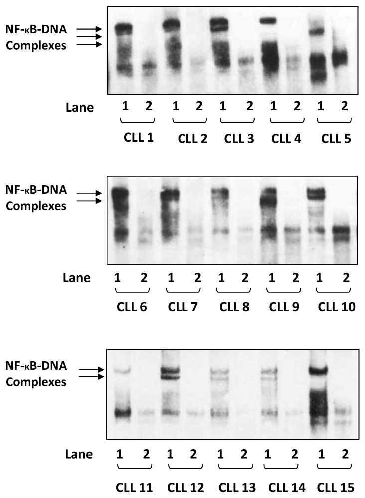
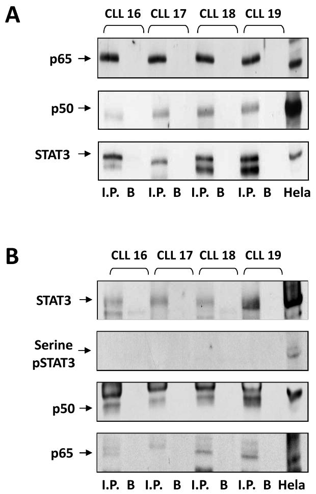
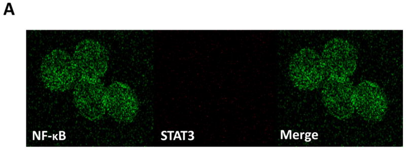
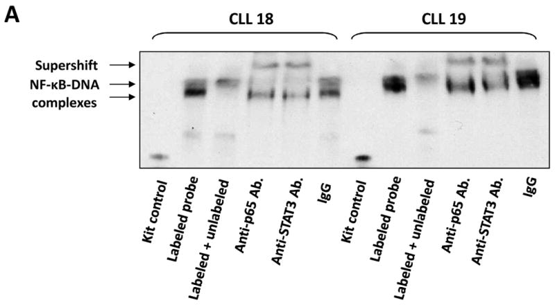
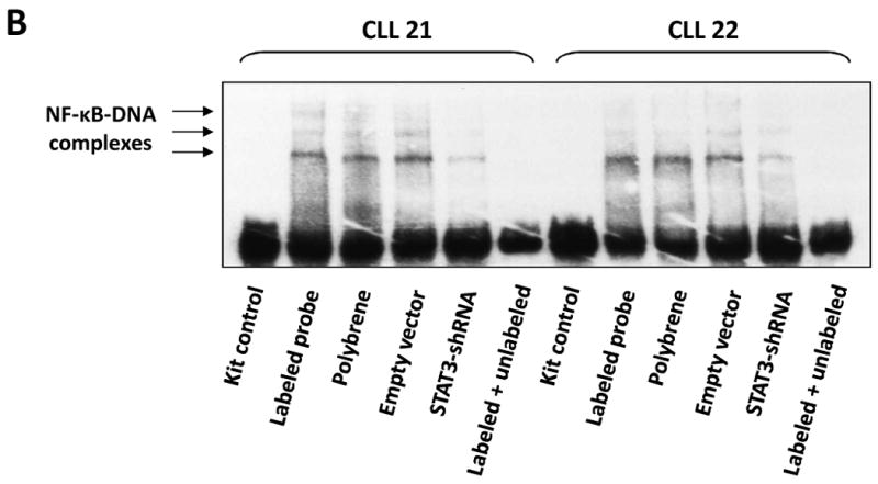
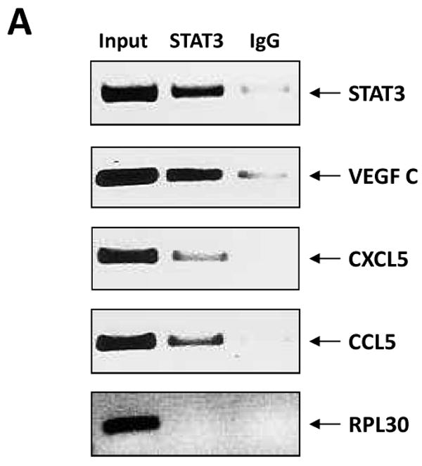
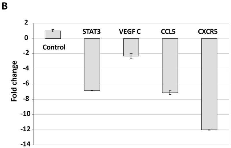
NF-κB plays a major role in the pathogenesis of B-cell neoplasms. A broad array of mostly extracellular stimuli has been reported to activate NF-κB, to various degrees, in chronic lymphocytic leukemia (CLL) cells. Because CLL cells harbor high levels of unphosphorylated STAT-3 (USTAT-3) and USTAT-3 was reported to activate NF-κB, we sought to determine whether USTAT-3 activates NF-κB in CLL. Using the electrophoretic mobility shift assay (EMSA), we studied peripheral blood low-density cells from 15 patients with CLL and found that CLL cell nuclear extracts from all the samples bound to an NF-κB DNA probe, suggesting that NF-κB is constitutively activated in CLL. Immunoprecipitation studies showed that STAT-3 bound NF-κB p65, and confocal microscopy studies detected USTAT-3/NF-κB complexes in the nuclei of CLL cells, thereby confirming these findings. Furthermore, infection of CLL cells with retroviral STAT-3-short hairpin RNA attenuated the binding of NF-κB to DNA, as assessed by EMSA, and downregulated mRNA levels of NF-κB-regulated genes, as assessed by quantitative PCR. Taken together, our data suggest that USTAT-3 binds to the NF-κB p50/p65 dimers and that the USTAT-3/NF-κB complexes bind to DNA and activate NF-κB-regulated genes in CLL cells.


Conflict of interest statement

Disclosure of potential Conflicts of Interest: The authors declare that no conflict of interest exists.

Figures

Figure 1
Figure 1
NF-κB is constitutively activated in CLL cells. EMSA studies of PB low-density cell nuclear extracts from 15 different patients with CLL are presented. Lane 1 of each sample depicts binding of CLL cell nuclear extract to a biotin-labeled (hot) NF-κB DNA probe, whereas lane 2 depicts a reduction in binding when an excess of unlabeled probe was added to the biotin-labeled (hot) κB DNA probe (hot + cold). In all 15 samples, NF-κB-DNA (p50/p65) complexes were detected and NF-κB-DNA binding was almost completely abolished by the cold probe.
Figure 2
Figure 2
U-STAT3 binds NF-κB p50/p65 dimers. A. PB low-density cell-protein extracts from four different patients with CLL were immunoprecipitated with anti-Stat3 antibodies. As shown, STAT3 and the NF-κB p65 and p50 proteins were detected in the immunoprecipitates of all three PB samples. B. PB low-density cell-protein extracts from the same patients with CLL were immunoprecipitated with anti-p65 antibodies. Hela cell protein extract was used as a positive control. As shown, p65, p50, STAT3, but not serine pSTAT3 proteins were detected in all immunopreciptates. I.P., immunoprecipitate; B, beads (control).
Figure 3
Figure 3
U-STAT3-NF-κB complexes are detected in CLL cells but not in normal CD19+ cells. A. Confocal microscopy studies of normal PB CD19+ cells detected a scattered signal of the NF-κB p65 protein in the cytoplasm and nucleus (left panel; green), faint signals of STAT3 (middle panel; red) but no signal of STAT3-p65 complexes (right panel; yellow). B. Confocal microscopy studies of PB CLL cells detected a dense signal of p65 (green), STAT3 (red), and STAT3-p65 complexes (yellow) in the cytoplasm and mainly in the nucleus. One field of CLL cells (upper panel) and a single CLL cell (lower panel) are shown. Depicted are representative photomicrographs (X 400) obtained from two different samples of normal CD19+ lymphocytes and CLL cells of patients 19 and 20.
Figure 3
Figure 3
U-STAT3-NF-κB complexes are detected in CLL cells but not in normal CD19+ cells. A. Confocal microscopy studies of normal PB CD19+ cells detected a scattered signal of the NF-κB p65 protein in the cytoplasm and nucleus (left panel; green), faint signals of STAT3 (middle panel; red) but no signal of STAT3-p65 complexes (right panel; yellow). B. Confocal microscopy studies of PB CLL cells detected a dense signal of p65 (green), STAT3 (red), and STAT3-p65 complexes (yellow) in the cytoplasm and mainly in the nucleus. One field of CLL cells (upper panel) and a single CLL cell (lower panel) are shown. Depicted are representative photomicrographs (X 400) obtained from two different samples of normal CD19+ lymphocytes and CLL cells of patients 19 and 20.
Figure 4
Figure 4
The U-STAT3/NF-κB complex binds to DNA. A. CLL cell nuclear proteins p65 and STAT3 bind to a biotinylated κB-DNA probe. EMSA of CLL cell nuclear extract is depicted. As shown, the addition of anti-p65 or anti-STAT3 antibodies to the κB DNA biotinylated probe induced a supershift, suggesting that p65 and STAT3 bind to DNA. B. STAT3-shRNA attenuates the binding of CLL cell nuclear extract to a biotin-labeled DNA probe. CLL cells were infected either with lentiviral STAT3-shRNA or empty virus. Nuclear protein was extracted and EMSA was performed. As shown, NF-κB-DNA complexes were detected, and excess unlabeled (cold) DNA probe abolished the binding. Furthermore, infections with STAT3-shRNA, but not with empty virus, significantly attenuated NF-κB-DNA binding.
Figure 4
Figure 4
The U-STAT3/NF-κB complex binds to DNA. A. CLL cell nuclear proteins p65 and STAT3 bind to a biotinylated κB-DNA probe. EMSA of CLL cell nuclear extract is depicted. As shown, the addition of anti-p65 or anti-STAT3 antibodies to the κB DNA biotinylated probe induced a supershift, suggesting that p65 and STAT3 bind to DNA. B. STAT3-shRNA attenuates the binding of CLL cell nuclear extract to a biotin-labeled DNA probe. CLL cells were infected either with lentiviral STAT3-shRNA or empty virus. Nuclear protein was extracted and EMSA was performed. As shown, NF-κB-DNA complexes were detected, and excess unlabeled (cold) DNA probe abolished the binding. Furthermore, infections with STAT3-shRNA, but not with empty virus, significantly attenuated NF-κB-DNA binding.
Figure 5
Figure 5
U-STAT3/NF-κB complexes activate NF-κB-target genes. A. STAT3-shRNA downregulated mRNA levels of specific NF-κB-regulated genes. As shown, infection with lentiviral STAT3-shRNA downregulated mRNA levels of VEGF C, CCL5, and CXCR5. As expected, lentiviral STAT3-shRNA downregulated the level of STAT3 mRNA as well. In this experiment we used PB cells from patient 21. B. STAT3 co-immunoprecipitates with DNA of NF-κB-regulated genes. CLL cell-derived chromatin was immunoprecipitated by anti-STAT3 antibodies or rabbit serum (IgG control). The co-immunoprecipitated DNA was amplified by PCR using promoter constructs of STAT3, VEGF C, CCL5, CXCR5, or the control gene RPL30. As shown, DNA of STAT3, VEGF C, CCL5 and CXCR5 but not the control RPL30 gene, co-immunoprecipitated with STAT3 protein. STAT3, VEGF C, CCL5, or RPL30 DNA was detected in whole cell chromatin-extracted DNA (Input).
Figure 5
Figure 5
U-STAT3/NF-κB complexes activate NF-κB-target genes. A. STAT3-shRNA downregulated mRNA levels of specific NF-κB-regulated genes. As shown, infection with lentiviral STAT3-shRNA downregulated mRNA levels of VEGF C, CCL5, and CXCR5. As expected, lentiviral STAT3-shRNA downregulated the level of STAT3 mRNA as well. In this experiment we used PB cells from patient 21. B. STAT3 co-immunoprecipitates with DNA of NF-κB-regulated genes. CLL cell-derived chromatin was immunoprecipitated by anti-STAT3 antibodies or rabbit serum (IgG control). The co-immunoprecipitated DNA was amplified by PCR using promoter constructs of STAT3, VEGF C, CCL5, CXCR5, or the control gene RPL30. As shown, DNA of STAT3, VEGF C, CCL5 and CXCR5 but not the control RPL30 gene, co-immunoprecipitated with STAT3 protein. STAT3, VEGF C, CCL5, or RPL30 DNA was detected in whole cell chromatin-extracted DNA (Input).

References

    1. Yee KW, O'Brien SM. Chronic lymphocytic leukemia: diagnosis and treatment. Mayo Clin Proc. 2006;81:1105–29. - PubMed
    1. Schuh K, Avots A, Tony HP, Serfling E, Kneitz C. Nuclear NF-ATp is a hallmark of unstimulated B cells from B-CLL patients. Leuk Lymphoma. 1996;23:583–92. - PubMed
    1. Sembries S, Pahl H, Stilgenbauer S, Dohner H, Schriever F. Reduced expression of adhesion molecules and cell signaling receptors by chronic lymphocytic leukemia cells with 11q deletion. Blood. 1999;93:624–31. - PubMed
    1. Furman RR, Asgary Z, Mascarenhas JO, Liou HC, Schattner EJ. Modulation of NF-kappa B activity and apoptosis in chronic lymphocytic leukemia B cells. J Immunol. 2000;164:2200–6. - PubMed
    1. Bernal A, Pastore RD, Asgary Z, et al. Survival of leukemic B cells promoted by engagement of the antigen receptor. Blood. 2001;98:3050–7. - PubMed

Publication types

MeSH terms