. 2011 Apr 28;117(17):4667-78.
doi: 10.1182/blood-2010-10-316356. Epub 2011 Mar 1.

An important role of lymphatic vessel activation in limiting acute inflammation

Affiliations

An important role of lymphatic vessel activation in limiting acute inflammation

Reto Huggenberger et al. Blood. .

Abstract

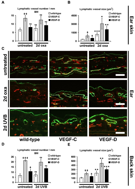
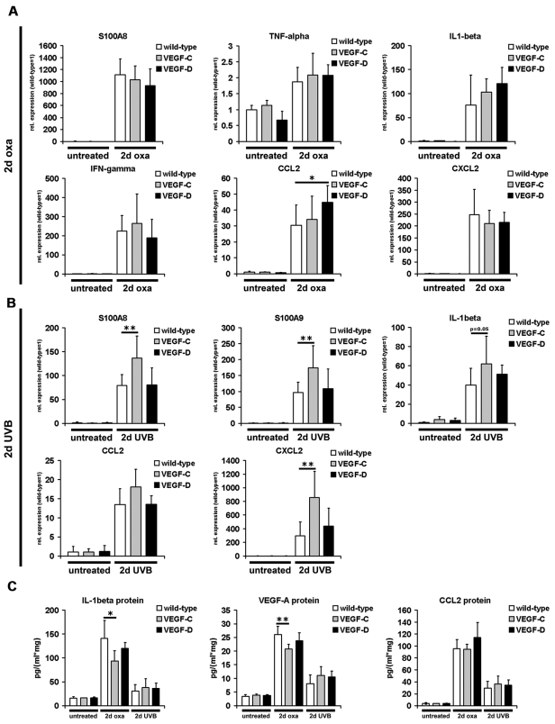
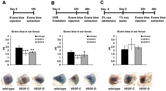
In contrast to the established role of blood vessel remodeling in inflammation, the biologic function of the lymphatic vasculature in acute inflammation has remained less explored. We studied 2 established models of acute cutaneous inflammation, namely, oxazolone-induced delayed-type hypersensitivity reactions and ultraviolet B irradiation, in keratin 14-vascular endothelial growth factor (VEGF)-C and keratin 14-VEGF-D transgenic mice. These mice have an expanded network of cutaneous lymphatic vessels. Transgenic delivery of the lymphangiogenic factors VEGF-C and the VEGFR-3 specific ligand mouse VEGF-D significantly limited acute skin inflammation in both experimental models, with a strong reduction of dermal edema. Expression of VEGFR-3 by lymphatic endothelium was strongly down-regulated at the mRNA and protein level in acutely inflamed skin, and no VEGFR-3 expression was detectable on inflamed blood vessels and dermal macrophages. There was no major change of the inflammatory cell infiltrate or the composition of the inflammatory cytokine milieu in the inflamed skin of VEGF-C or VEGF-D transgenic mice. However, the increased network of lymphatic vessels in these mice significantly enhanced lymphatic drainage from the ear skin. These results provide evidence that specific lymphatic vessel activation limits acute skin inflammation via promotion of lymph flow from the skin and reduction of edema formation.


Figures

Figure 1
Figure 1
Lymphatic vessel activation reduces edema during acute skin inflammation. (A) K14-VEGF-C (●, n = 7), K14-VEGF-D (●, n = 8) Tg mice, and their wild-type littermates (□, n = 12) were painted with 2% oxazolone and challenged, 5 days later, with 1% oxazolone on the ears. The ear thickness of K14-VEGF-C and K14-VEGF-D Tg mice was significantly reduced compared with wild-type controls at the indicated time points. (B) K14-VEGF-C (●, n = 7), K14-VEGF-D (●, n = 5) Tg mice, and their wild-type littermates (□, n = 11) were irradiated with 200 mJ/cm2 UVB light, and the ear thickness was measured using calipers. The ear thickness of K14-VEGF-C and K14-VEGF-D Tg mice was significantly reduced compared with wild-type controls until day 8 after UVB irradiation. (A-B) Data are mean ± SEM. *P ≤ .05. **P ≤ .01. ***P ≤ .001. (C-E) Hematoxylin and eosin stains of untreated mouse ears (C), at day 2 after oxazolone challenge (2-day oxa, D) or UVB irradiation (2-day UVB, E) revealed reduced edema in inflamed K14-VEGF-C and K14-VEGF-D Tg mice, compared with inflamed skin of wild-type mice. One ear-half is shown. Bars represent 100 μm.
Figure 2
Figure 2
Enlargement of cutaneous lymphatic vessels during acute inflammation. (A-B) Quantitative image analyses of CD31+/LYVE-1+ lymphatic vessels in the ear skin of mice revealed a significantly increased number per millimeter basement membrane (BM, A) and size (B) of lymphatic vessels in untreated K14-VEGF-C Tg mice, compared with untreated wild-type mice (n = 3 mice per group). The lymphatic vessel size was also increased in untreated K14-VEGF-D Tg mice, compared with untreated wild-type mice (n = 3 mice per group). The average number of lymphatic vessels in wild-type mice was not significantly different at 2 days after oxazolone challenge (2-day oxa) compared with untreated wild-type mice. The lymphatic vessel number was also not significantly different between untreated and oxazolone-challenged K14-VEGF-C and K14-VEGF-D Tg mice, respectively (A; wild-type, n = 10; K14-VEGF-C, n = 5; K14-VEGF-D, n = 5). At 2 days after oxazolone challenge, the average size of lymphatic vessels was significantly increased in K14-VEGF-C, K14-VEGF-D Tg, and wild-type mice, compared with untreated mice of the same genotype (B). (C) Representative images of CD31+/LYVE-1+ lymphatic vessels (green) in the ear skin. CD31+/LYVE-1 structures represent blood vessels (red). The positive staining of LYVE-1 in the stratum corneum in panel C is unspecific. Bars represent 100 μm. (D-E) The average lymphatic vessel number (D) and size (E) were significantly increased in the back skin of untreated K14-VEGF-C and K14-VEGF-D Tg mice, compared with untreated wild-type mice. At 2 days after UVB irradiation (2-day UVB), lymphatic vessel size (E), but not lymphatic vessel number (D), was significantly increased in K14-VEGF-C, K14-VEGF-D, and wild-type mice compared with untreated mice of the same genotype. (A-B,D-E) Data are mean ± SD. ‡P ≤ .05. ‡‡P ≤ .01. ‡‡‡P ≤ .001. ns indicates not significant versus untreated wild-type. *P ≤ .05. **P ≤ .01. ***P ≤ .001. ns indicates not significant versus untreated mice (untreated wild-type vs 2-day oxa/2-day UVB wild-type; untreated VEGF-C vs 2-day oxa/2-day UVB VEGF-C; untreated VEGF-D vs 2-day oxa/2-day UVB VEGF-D).
Figure 3
Figure 3
Enlargement of blood vessels during acute skin inflammation. (A-B) Immunofluorescence analyses for the blood vessel-specific marker MECA-32 and subsequent morphometric quantification showed a significant increase in blood vessel number per millimeter basement membrane (BM) in K14-VEGF-C and K14-VEGF-D Tg mice at 2 days after oxazolone challenge (2-day oxa), compared with untreated mice (A). The blood vessel size was also increased in all 3 groups of mice at 2 days of oxazolone challenge, compared with untreated mice of the same genotype (B). There was no significant difference in blood vessel number (A,C) or blood vessel size (B,D) at 2 days after oxazolone challenge or UVB irradiation (2-day UVB) between K14-VEGF-C, K14-VEGF-D Tg, and wild-type mice. (A-B) Quantification of ear skin. (C-D) Quantification of back skin. (A-D) Data are mean ± SD. *P ≤ .05. **P ≤ .01. ***P ≤ .001. ns indicates not significant versus untreated mice of the same genotype.
Figure 4
Figure 4
Down-regulation of VEGFR-3 during acute skin inflammation. (A-B) TaqMan-based real-time RT-PCR analyses were performed on whole ear and back skin extracts after 2 days of oxazolone challenge (2-day oxa; n = 6-10 per group) or UVB irradiation (2-day UVB; n = 5-8 per group) and in untreated K14-VEGF-C, K14-VEGF-D Tg, and wild-type mice (n = 3-5 per group). VEGFR-3 was significantly up-regulated in untreated K14-VEGF-C and K14-VEGF-D Tg mouse ear (A) and back skin (B) compared with untreated wild-type mice. VEGFR-3 was significantly down-regulated at 2 days of oxazolone challenge (A) or UVB irradiation (B) in all 3 groups of mice compared with untreated mice of the same genotype. (C-D) Single-cell suspensions from the ear of normal and oxazolone challenged mice were analyzed by FACS. (C) CD31+/CD45 cells represent endothelial cells, whereas CD31+/podoplanin+/CD45 cells are lymphatic endothelial cells. (D) Cutaneous lymphatic endothelial cells from inflamed ears of wild-type mice (2 days after oxazolone challenge) showed a 5-fold decrease of VEGFR-3 mRNA transcript levels compared with lymphatic endothelial cells from untreated mice. (A-B,D) Data are mean ± SD. ‡‡‡P ≤ .001 versus untreated wild-type. ***P ≤ .001 versus untreated mice of the same genotype. (E-F) Double immunofluorescence analyses of VEGFR-3 (red) and LYVE-1 (green) stains demonstrated that VEGFR-3 was strongly down-regulated on LYVE-1+ lymphatic vessels at 2 days after oxazolone challenge (E) or UVB irradiation (F) in the ear and back skin of wild-type mice (arrows). The positive staining of LYVE-1 in the stratum corneum (E) is unspecific. Bars represent 100 μm.
Figure 5
Figure 5
Inflammation marker expression in the skin during acute inflammation. (A-B) Real-time RT-PCR analyses were performed using RNAs from whole ear and back skin harvested 2 days after oxazolone challenge (2-day oxa; n = 6-10 per group) or UVB irradiation (2-day UVB; n = 5-8 per group) and from skin of untreated K14-VEGF-C, K14-VEGF-D Tg, and wild-type mice (n = 3-5 per group). All inflammation markers shown were significantly up-regulated in K14-VEGF-C, K14-VEGF-D Tg, and wild-type mouse ear (A) and back skin (B) 2 days after oxazolone challenge (A) or UVB irradiation (B), compared with untreated wild-type or transgenic mice. S100A8, S100A9, and CXCL2 mRNA levels were slightly increased in K14-VEGF-C Tg mice 2 days after UVB irradiation compared with irradiated wild-type mice (B). (C) ELISA analyses of ear lysates showed significantly increased levels of IL-1β, VEGF-A, and CCL2 at 2 days after oxazolone challenge or UVB irradiation in the ear skin of K14-VEGF-C, K14-VEGF-D Tg, and wild-type mice compared with untreated mice. The protein levels of IL-1β and VEGF-A were slightly but significantly reduced in the ear skin of oxazolone challenged K14-VEGF-C Tg mice compared with oxazolone challenged wild-type mice. (A-C) Data are mean ± SD. *P ≤ .05 versus oxazolone challenged or UVB irradiated wild-type mice. **P ≤ .01 versus oxazolone challenged or UVB irradiated wild-type mice.
Figure 6
Figure 6
Inflammatory cell infiltration during acute skin inflammation. (A-D) Immunofluorescence analyses for the monocyte/granulocyte marker CD11b and subsequent computer-based quantification showed a significantly increased number of dermal CD11b+ cells per millimeter basement membrane (BM) 2 days after oxazolone challenge (2 days oxa, A,C; ear skin) or UVB irradiation (2 days UVB, B,D; back skin) in K14-VEGF-C, K14-VEGF-D Tg, and wild-type mice, compared with untreated mice of the same genotype. K14-VEGF-C Tg mice had even more infiltrated dermal CD11b+ cells in their ear skin 2 days after oxazolone challenge compared with oxazolone challenged wild-type mice (A,C). The hair follicle sebaceous glands are stained red (C-D) because of endogenous biotin. (A-D) Data are mean ± SD. ***P ≤ .001. ns indicates not significant versus oxazolone challenged or UVB-irradiated wild-type mice. Bars represent 100 μm (C), 50 μm (D).
Figure 7
Figure 7
Increased lymph flow from the ear in K14-VEGF-C and K14-VEGF-D Tg mice. (A-C) Evans blue dye was intradermally injected into the ear of untreated or inflamed K14-VEGF-C, K14-VEGF-D Tg, and wild-type mice and was extracted 16 hours after the dye injection. The total dye remaining in the ear skin is indicated. Representative pictures before Evans blue extraction are shown at the bottom. (A) Untreated K14-VEGF-C (n = 5) and K14-VEGF-D (n = 5) Tg mice had significantly less Evans blue remaining in their ear skin 16 hours after injection compared with wild-type mice (n = 10). (B) Evans blue was also injected 32 hours after UVB irradiation and extracted 16 hours later. UVB irradiated K14-VEGF-C (n = 5) and K14-VEGF-D (n = 4) Tg mice showed a faster lymph flow than irradiated wild-type mice (n = 10). (C) There was no difference in Evans blue clearance from the inflamed ear during acute inflammation after oxazolone challenge between all mice (wild-type, n = 10; K14-VEGF-C, n = 5; K14-VEGF-D, n = 5). Data are mean ± SD. *P ≤ .05 versus wild-type mice. **P ≤ .01 versus wild-type mice. ***P ≤ .001 versus wild-type mice.

References

    1. Halin C, Detmar M. Inflammation, angiogenesis, and lymphangiogenesis. Methods Enzymol. 2008;445:1–25. - PubMed
    1. Bainbridge J, Sivakumar B, Paleolog E. Angiogenesis as a therapeutic target in arthritis: lessons from oncology. Curr Pharm Des. 2006;12(21):2631–2644. - PubMed
    1. Baluk P, Tammela T, Ator E, et al. Pathogenesis of persistent lymphatic vessel hyperplasia in chronic airway inflammation. J Clin Invest. 2005;115(2):247–257. - PMC - PubMed
    1. Danese S, Sans M, de la Motte C, et al. Angiogenesis as a novel component of inflammatory bowel disease pathogenesis. Gastroenterology. 2006;130(7):2060–2073. - PubMed
    1. Detmar M, Brown LF, Claffey KP, et al. Overexpression of vascular permeability factor/vascular endothelial growth factor and its receptors in psoriasis. J Exp Med. 1994;180(3):1141–1146. - PMC - PubMed

MeSH terms