. 2011 Apr;121(4):1386-96.
doi: 10.1172/JCI44883.

The liver-specific microRNA miR-122 controls systemic iron homeostasis in mice

Affiliations

The liver-specific microRNA miR-122 controls systemic iron homeostasis in mice

Mirco Castoldi et al. J Clin Invest. 2011 Apr.

Abstract

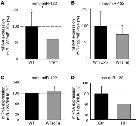
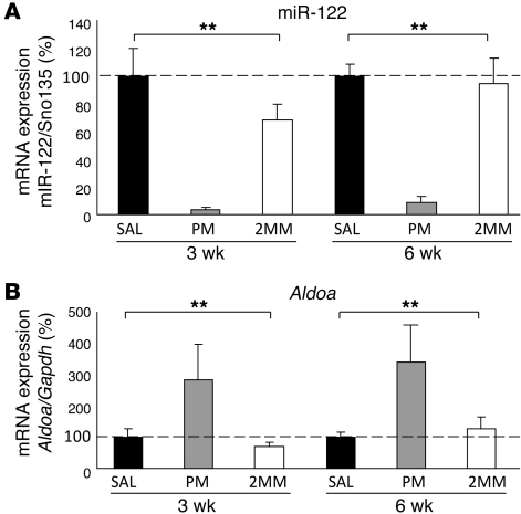
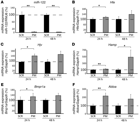
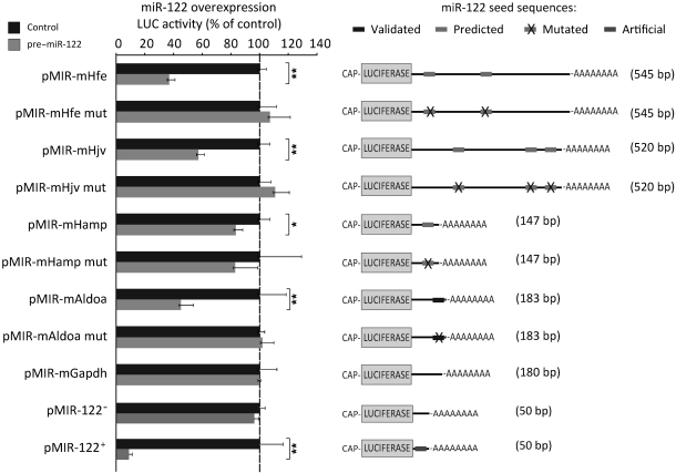
Systemic iron homeostasis is mainly controlled by the liver through synthesis of the peptide hormone hepcidin (encoded by Hamp), the key regulator of duodenal iron absorption and macrophage iron release. Here we show that the liver-specific microRNA miR-122 is important for regulating Hamp mRNA expression and tissue iron levels. Efficient and specific depletion of miR-122 by injection of a locked-nucleic-acid-modified (LNA-modified) anti-miR into WT mice caused systemic iron deficiency, characterized by reduced plasma and liver iron levels, mildly impaired hematopoiesis, and increased extramedullary erythropoiesis in the spleen. Moreover, miR-122 inhibition increased the amount of mRNA transcribed by genes that control systemic iron levels, such as hemochromatosis (Hfe), hemojuvelin (Hjv), bone morphogenetic protein receptor type 1A (Bmpr1a), and Hamp. Importantly, miR-122 directly targeted the 3′ untranslated region of 2 mRNAs that encode activators of hepcidin expression, Hfe and Hjv. These data help to explain the increased Hamp mRNA levels and subsequent iron deficiency in mice with reduced miR-122 levels and establish a direct mechanistic link between miR-122 and the regulation of systemic iron metabolism.


Figures

Figure 1
Figure 1. miR-122 levels are decreased in Hfe–/– mice and patients with HH.
(A) qPCR analysis of miR-122 expression in liver total RNA of WT (n = 8) and Hfe–/– (n = 11) mice (P = 0.039). mmu-miR-122, Mus musculus miR-122 stem-loop. (B) Analysis of miR-122 expression in the liver of WT mice injected with dextran (Dxt; n = 5) and iron-dextran (Fe; n = 6) (P = 0.187) and (C) WT mice on a regular (n = 4) or iron-supplemented diet (dFe; n = 4) (P = 0.471). (D) miR-122 levels were reduced in liver biopsies of HH patients (n = 6) compared with the control group without iron overload (Ctr; n = 4) (P = 0.068). hsa-miR-122, Homo sapiens miR-122 stem-loop. Data were normalized to the appropriate reference genes: miR-194 (A and B), mouse RNU6 (C), and human RNU6 (D). Data are mean ± SD, and values from WT mouse (AC) and control patient (D) groups were set to 100%. *P < 0.05, 2-tailed Student’s t test.
Figure 2
Figure 2. miR-122 depletion is functional.
(A) miR-122 detectability was decreased in the liver of PM_anti–miR-122–injected mice. Mice were injected i.p. with a single dose of 25 μg/g PM_anti–miR-122 (PM), 2MM_anti–miR-122 (2MM), or saline (SAL) and sacrificed 3 or 6 weeks after injection. Hepatic miR-122 expression was analyzed by qPCR, and data were normalized to the expression of small nucleolar RNA Sno135. Detectability of miR-122 was reduced 3 (P = 0.0001) and 6 (P = 0.0001) weeks after PM_anti–miR-122 administration. (B) Aldoa was increased in the liver of PM_anti–miR-122–injected mice 3 (P = 0.0001) and 6 (P = 0.0001) weeks after treatment. Values were normalized to mRNA expression of reference gene Gapdh. Data are mean ± SD (n = 8), and the saline-treated group was set to 100%. **P < 0.01, 1-way ANOVA.
Figure 3
Figure 3. Liver mRNA expression of Hamp, Hfe, Hjv, and Bmpr1a is increased in PM_anti–miR-122–treated mice.
Analysis of hepatic (A) Hamp (3 weeks, P = 0.0005; 6 weeks, P = 0.27), (B) Hfe (3 weeks, P = 0.0005; 6 weeks, P = 0.158), (C) Hjv (3 weeks, P = 0.0001; 6 weeks, P = 0.0225), and (D) Bmpr1a (3 weeks, P = 0.0001; 6 weeks, P = 0.011) mRNA expression by qPCR 3 and 6 weeks after anti-miR administration. Values were normalized to mRNA expression of reference gene Gapdh. Data are mean ± SD (n = 8), and the saline-treated group was set to 100%. *P < 0.05, **P < 0.01, 1-way ANOVA.
Figure 4
Figure 4. miR-122 inhibition in murine primary hepatocytes.
Primary hepatocytes were transfected with PM_anti–miR-122 or scrambled control oligos (SCR). (A) miR-122 expression was analyzed 24 (P = 0.01) and 48 (P = 0.01) hours after transfection. (BF) Expression of (B) Hfe (24 hours, P = 0.026; 48 hours, P = 0.117), (C) Hjv (24 hours, P = 0.014; 48 hours, P = 0.11), (D) Hamp (24 hours, P = 0.005; 48 hours, P = 0.044), (E) Bmpr1a (24 hours, P = 0.034; 48 hours, P = 0.175), and (F) Aldoa (24 hours, P = 0.003; 48 hours, P = 0.119) mRNA 24 and 48 hours after transfection of PM_anti–miR-122 or scrambled control oligos. Data are mean ± SD (n = 4), and the value for scrambled control–transfected cells was set to 100%. Data were normalized to mRNA expression of the appropriate reference gene. *P < 0.05, **P < 0.01, 2-tailed Student’s t test.
Figure 5
Figure 5. Validation of Hfe and Hjv as miR-122 target genes.
Hepa1-6 cells were transfected with pMIR-mHfe, pMIR-mHjv, pMIR-mHamp, pMIR-mAldoa, or pMIR-mGapdh (see Supplemental Table 3 for localization of predicted miR-122 binding sites), miR-122 complementary sequence (pMIR-122+), or miR-122 antisense sequence (pMIR-122), either with (pre–miR-122) or without (control) miR-122 mimics. Luciferase activity was measured 24 hours later. As a specificity control, we transfected luciferase reporter vectors linked to the 3′-UTRs of the genes of interest that contain mutations within the predicted miR-122 seed sequences (mut; see Supplemental Table 3). Experiments were performed at least in triplicate, and results of the luciferase reporter assay are presented as fold change ± SD of the pre–miR-122– and control-transfected cells. *P < 0.05, **P < 0.01, 2-tailed Student’s t test.
Figure 6
Figure 6. Extramedullary hematopoiesis in PM_anti–miR-122–injected mice.
(A) Heat map representing genes with increased mRNA expression in the spleen of PM_anti–miR-122 treated mice, as assessed by IronChip. n.c., not changed. (B) Splenic Tfr1 (P = 0.007), Hmbs (P = 0.08), Hba-a1 (P = 0.24), and Alas2 (P = 0.14) mRNA expression, as assessed by qPCR 3 weeks after injection of anti-miRs. Data were normalized to mRNA expression of reference gene Actb. (C) Splenic expression of miR-451 (3 weeks, P = 0.0016; 6 weeks, P = 0.979), miR-29b (3 weeks, P = 0.0019; 6 weeks, P = 0.844), and miR-17 (3 weeks, P = 0.093; 6 weeks, P = 0.19) by miRNA qPCR 3 and 6 weeks after injection of anti-miRs. Data were normalized to expression of miR-452, which remained unchanged in miRNA expression profiles of splenic total RNAs of the same mice. (D) Splenic Gata1 (P = 0.003) and Gata2 (P = 0.041) mRNA expression 3 weeks after injection of anti-miRs. Data are mean ± SD (n = 8), and values from the saline-treated group were set to 100%. *P < 0.05, **P < 0.01; 1-way ANOVA.

Comment in

References

    1. Hentze MW, Muckenthaler MU, Galy B, Camaschella C. Two to tango: regulation of Mammalian iron metabolism. Cell. 2010;142(1):24–38. doi: 10.1016/j.cell.2010.06.028. - DOI - PubMed
    1. Krause A, et al. LEAP-1, a novel highly disulfide-bonded human peptide, exhibits antimicrobial activity. FEBS Lett. 2000;480(2–3):147–150. doi: 10.1016/S0014-5793(00)01920-7. - DOI - PubMed
    1. Park CH, Valore EV, Waring AJ, Ganz T. Hepcidin, a urinary antimicrobial peptide synthesized in the liver. J Biol Chem. 2001;276(11):7806–7810. doi: 10.1074/jbc.M008922200. - DOI - PubMed
    1. Nemeth E, et al. Hepcidin regulates cellular iron efflux by binding to ferroportin and inducing its internalization. Science. 2004;306(5704):2090–2093. doi: 10.1126/science.1104742. - DOI - PubMed
    1. Finberg KE, et al. Mutations in TMPRSS6 cause iron-refractory iron deficiency anemia (IRIDA). Nat Genet. 2008;40(5):569–571. doi: 10.1038/ng.130. - DOI - PMC - PubMed

Publication types

MeSH terms