. 2011 Apr;121(4):1636-45.
doi: 10.1172/JCI42953.

PML-RARA can increase hematopoietic self-renewal without causing a myeloproliferative disease in mice

Affiliations

PML-RARA can increase hematopoietic self-renewal without causing a myeloproliferative disease in mice

John S Welch et al. J Clin Invest. 2011 Apr.

Abstract

Acute promyelocytic leukemia (APL) is characterized by the t(15;17) translocation that generates the fusion protein promyelocytic leukemia-retinoic acid receptor α (PML-RARA) in nearly all cases. Multiple prior mouse models of APL constitutively express PML-RARA from a variety of non-Pml loci. Typically, all animals develop a myeloproliferative disease, followed by leukemia in a subset of animals after a long latent period. In contrast, human APL is not associated with an antecedent stage of myeloproliferation. To address this discrepancy, we have generated a system whereby PML-RARA expression is somatically acquired from the mouse Pml locus in the context of Pml haploinsufficiency. We found that physiologic PML-RARA expression was sufficient to direct a hematopoietic progenitor self-renewal program in vitro and in vivo. However, this expansion was not associated with evidence of myeloproliferation, more accurately reflecting the clinical presentation of human APL. Thus, at physiologic doses, PML-RARA primarily acts to increase hematopoietic progenitor self-renewal, expanding a population of cells that are susceptible to acquiring secondary mutations that cause progression to leukemia. This mouse model provides a platform for more accurately dissecting the early events in APL pathogenesis.


Figures
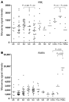
Figure 1
Figure 1. Expression profiling.
(A and B) Expression array profile of (A) human PML and (B) human RARA in 111 patients with indicated FAB-AML and 5 flow-sorted normal controls. CD34, CD34+ bone marrow cells; Pros, promyelocytes. Individual data points represent results from individual patients. Horizontal bars are the median value.
Figure 2
Figure 2. mPML-PRflox targeting strategy and the result of intercrosses with CMV-Cre, LysM-Cre, and ERT2-Cre mice.
(A) Targeting strategy. Rectangles represent exons. (B) The results of intercrosses with mPML-PRflox × CMV-Cre, (C) mPML-PRflox × LysM-Cre, and (D) mPML-PRflox × ERT2-Cre mice are shown, with details about resulting pups.
Figure 3
Figure 3. mPML-fPR does not grossly alter bone marrow progenitor populations, CFUs, or ex vivo myeloid differentiation.
(A) Schema showing that mice were treated with 10 doses of tamoxifen (4 mg i.p. twice weekly for 5 weeks) and analyzed 2 weeks later. (B) mPML-fPR modestly decreased the percentage of LincKit+Sca+ (KLS) cells but not the percentage of LincKit+ScaCD16/32+CD34+ (GMP), LincKit+ScaCD16/32CD34+ (CMP), or LincKit+ScaCD16/32CD34 (MEP) cells. KLS is expressed as the percentage of Lin cells; the others are expressed as the percentage of LincKit+Sca cells. Results are from 4 independent mice in each genotype. Horizontal bars are the median value. (C) mPML-fPR did not alter the number of CFUs in the bone marrow cells when grown for 7 days in methylcellulose containing IL-3, IL-6, and SCF (average ± SD). (D) mPML-fPR did not alter the immunophenotype of cells maturing during the methylcellulose culture in C. After 1 week in culture, cells were collected from methylcellulose, washed, and stained as indicated. Representative results are from 2 experiments (average ± SD). Horizontal bars are the median value. (E) Acute exposure of ERT2-Cre+/– × mPML-PRflox+/– bone marrow cells to 4-hydroxytamoxifen (4-OH tam) (0.1 M or 1 M) modestly decreased the number of CFU-Ms and total CFUs. Bone marrow cells from mice without prior tamoxifen exposure were harvested and plated in methylcellulose, as above, containing increasing doses of 4-hydroxytamoxifen (average ± SD).
Figure 4
Figure 4. mPML-fPR leads to inappropriate ex vivo self-renewal of bone marrow CFUs.
(A) Experiment schema showing that ERT2-Cre+/– × mPML-PRflox+/– mice and littermate controls were treated with tamoxifen (4 mg i.p. twice weekly for 10 doses). Bone marrow cells were plated in methylcellulose containing IL-3, IL-6, and SCF. At 7-day intervals, colony numbers were counted and cells were harvested and replated as indicated. (B) mPML-PRflox leads to inappropriate self-renewal when intercrossed with ERT2-Cre but not LysM-Cre. Representative results are from 2 experiments. Data points represent average and SD from 4 individual mice. (C) The mPML-fPR allele was associated with continued CFU activity in ERT2-Cre, but not LysM-Cre, bone marrow. The percentage of mPML-fPR alleles was assessed at each time point in B using qPCR. Data points represent results from individual mice. Horizontal bars are the median value.
Figure 5
Figure 5. Rare leukemia in LysM-Cre × mPML-PRflox mice.
(A) A tumor watch of LysM-Cre+/– × mPML-PRflox+/– mice (n = 54) resulted in leukemic mouse (mouse 6317, founder line 51) with 90% and 95% peripheral blood and spleen mPML-fPR alleles, respectively. Nonleukemic mice were evaluated at 18 months and showed little evidence of expanded mPML-fPR alleles in either peripheral blood or spleen. (B) Spleen cell cytomorphology from mouse 6317, stained with Wright-Giemsa (original magnification, ×1,000). (CE) Spleen cells from mouse 6317 were transplanted into nonirradiated B6 mice (n = 5). In 6 weeks all mice were moribund, with (C) elevated white blood cell counts and (D) splenomegaly. (E) qPCR evaluation of the spleen cells in moribund secondary recipients revealed 100% mPML-fPR. (F and G) Analysis of LysM-Cre+/– × mPML-PRflox+/– mice and littermate controls at 18 months revealed normal (F) white blood cell and (G) spleen sizes, with rare evidence of splenomegaly in LysM-Cre+/– mice, regardless of mPML-PRflox genotype. Data points represent results from individual mice. Horizontal bars are the median value.
Figure 6
Figure 6. Expansion of mPML-fPR alleles after pulsed tamoxifen.
(A) Experimental schema showing ERT2-Cre+/– × mPML-PRflox+/– mice and mPML-PRflox+/– littermate controls were treated with 0, 1, or 5 doses of tamoxifen (4 mg i.p.). (B) mPML-fPR did not alter peripheral blood counts at 6 months. NE, neutrophil; Ly, lymphocytes; Mo, monocytes; Eo, eosinophils; Hgb, hemoglobin; Plt, platelet. (C and D) After the initial pulse of tamoxifen, the percentage of mPML-fPR alleles in peripheral blood increased and was measured using qPCR at indicated time points. Numbers next to colored dots indicate specific mice. (E) At 18 months, the percentage of mPML-fPR alleles was assessed in bone marrow cells. (F) Expansion of mPML-fPR alleles was not associated with splenomegaly. Data represent results from individual mice at indicated time points. Horizontal bars are the median value.
Figure 7
Figure 7. Competitive and noncompetitive transplantation.
Bone marrow from indicated donor mice (CD45.2) was transplanted with competitor bone marrow (CD45.1) into lethally irradiated recipients (CD45.1). After engraftment, recipients were treated with the indicated doses of tamoxifen (3 mg by gavage). At the indicated time points, peripheral blood (PB) was assessed by flow cytometry. (A) Assessment of ERT2-Cre+/– × mPML-PRflox+/– donor bone marrow cells. (B) Assessment of ERT2-Cre+/– donor bone marrow cells. (C) Assessment of ERT2-Cre+/– × mPML-PRflox+/– donor bone marrow cells. Results represent average and SD from indicated numbers of mice in each cohort.

References

    1. Jurcic JG, Soignet SL, Maslak AP. Diagnosis and treatment of acute promyelocytic leukemia. Curr Oncol Rep. 2007;9(5):337–344. doi: 10.1007/s11912-007-0045-9. - DOI - PubMed
    1. Borrow J, et al. Diagnosis of acute promyelocytic leukaemia by RT-PCR: detection of PML-RARA and RARA-PML fusion transcripts. Br J Haematol. 1992;82(3):529–540. doi: 10.1111/j.1365-2141.1992.tb06463.x. - DOI - PubMed
    1. Kogan SC. Mouse models of acute promyelocytic leukemia. Curr Top Microbiol Immunol. 2007;313:3–29. - PubMed
    1. Westervelt P, Ley TJ. Seed versus soil: the importance of the target cell for transgenic models of human leukemias. Blood. 1999;93(7):2143–2148. - PubMed
    1. Grignani F, et al. PML/RAR alpha fusion protein expression in normal human hematopoietic progenitors dictates myeloid commitment and the promyelocytic phenotype. Blood. 2000;96(4):1531–1537. - PubMed

Publication types

MeSH terms

Substances