. 2011 Apr;121(4):1585-95.
doi: 10.1172/JCI44630.

A Tbx1-Six1/Eya1-Fgf8 genetic pathway controls mammalian cardiovascular and craniofacial morphogenesis

Affiliations

A Tbx1-Six1/Eya1-Fgf8 genetic pathway controls mammalian cardiovascular and craniofacial morphogenesis

Chaoshe Guo et al. J Clin Invest. 2011 Apr.

Erratum in

  • J Clin Invest. 2011 May 2;121(5):2060

Abstract

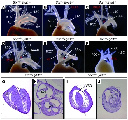
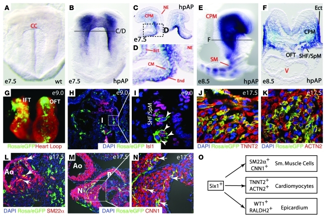
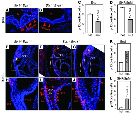
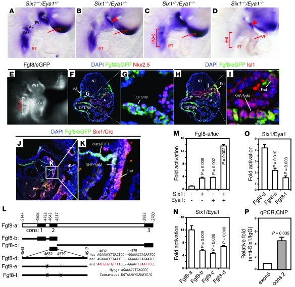
Shared molecular programs govern the formation of heart and head during mammalian embryogenesis. Development of both structures is disrupted in human chromosomal microdeletion of 22q11.2 (del22q11), which causes DiGeorge syndrome (DGS) and velo-cardio-facial syndrome (VCFS). Here, we have identified a genetic pathway involving the Six1/Eya1 transcription complex that regulates cardiovascular and craniofacial development. We demonstrate that murine mutation of both Six1 and Eya1 recapitulated most features of human del22q11 syndromes, including craniofacial, cardiac outflow tract, and aortic arch malformations. The mutant phenotypes were attributable in part to a reduction of fibroblast growth factor 8 (Fgf8), which was shown to be a direct downstream effector of Six1 and Eya1. Furthermore, we showed that Six1 and Eya1 genetically interacted with Fgf8 and the critical del22q11 gene T-box transcription factor 1 (Tbx1) in mice. Together, these findings reveal a Tbx1-Six1/Eya1-Fgf8 genetic pathway that is crucial for mammalian cardiocraniofacial morphogenesis and provide insights into the pathogenesis of human del22q11 syndromes.


Figures

Figure 1
Figure 1. Six1–/–Eya1–/– mutants phenocopy features of human del22q11 syndromes.
(AF) Gross morphological defects of the OFT and great arteries of newborn mutants (BF) versus WT control (A). (GJ) H&E histological staining of sectioned hearts revealed VSD and dysmorphology of the outflow valves in a Six1–/–Eya1–/– mutant (I and J) compared with Six1+/–Eya1+/– control (G and H). Ao, aortic artery; BCA, brachiocephalic artery; dA, descending aorta; DLCC, duplicated left common carotid artery; LCC, left common carotid artery; LSC, left subclavian artery; p, pulmonary artery; RCC, right common carotid artery; RRSA, retroesophageal right subclavian artery; RSC, right subclavian artery; V, ventricle; VR, vascular ring. Asterisks denote interrupted aortic arches. Original magnification, ×50 (G and I); ×80 (H and J).
Figure 2
Figure 2. Six1 is expressed in cardiac progenitors.
(AF) Six1 is expressed in cardiac/skeletal muscle progenitors and in cranial ectoderm and endoderm at E7.5 and E8.5. WT (A) and Six1Cre/hpAP (BF) embryos were stained for hpAP activity. Cross-sections of B are shown in C and D; cross-section of E is shown in F. CC, cardiac crescent; Ect, ectoderm; End, endoderm; NE, neural epithelium; SM, somite. (G) Whole-mount dorsal-right view of the looped heart from an E9.0 Six1Cre/hpAPR26RmTmG embryo. eGFP+ cells (Rosa/eGFP) in the Six1 lineage were found in inflow tract (IFT), OFT, and developing apex. (H and I) eGFP+ cells in the Six1 lineage and Isl1 colabeled SHF cardiac progenitor cells (arrowheads) in E9.0 sagittal sectioned tissue. Boxed region in H is enlarged in I. (JN) Coimmunostaining of eGFP+ cells in the Six1 lineage with TNNT2 (J) and ACTN2 (K) in the RV and with SM22α (L) and CNN1 (M and N, arrows) in the proximal region of the great arteries from E17.5 hearts. Boxed region in M is enlarged in N. Original magnification, ×115 (A and B); ×100 (C, D, and F); ×63 (E); ×200 (GN). (O) Summary of Six1+ progenitor cell lineage, based on results from Figure 2 and Supplemental Figure 5 (WT1, RALDH2; epicardial progenitor data).
Figure 3
Figure 3. Six1 and Eya1 are required for cell proliferation and survival in PA and OFT.
(AD) Anti-pH3 immunostaining (red) of E9.5 transverse sections to label proliferating cells. Quantification of results are shown in C and D. FG, foregut; mut, Six1–/–Eya1–/– mutant; het, heterozygous control. n = 3. (EL) TUNEL staining of sagittal sections revealed increased cell death in pharyngeal endoderm, SHF/SpM, and pharyngeal mesenchyme (PM) that contained CNCs and mesoderm. Boxed regions in EG are enlarged in HJ. Original magnification, ×200 (A, B, and EJ). n = 3. P values were determined by Student’s t test.
Figure 4
Figure 4. Six1 and Eya1 directly regulate Fgf8 expression.
(AD) Fgf8 expression was dependent on Six1 and Eya1. Fgf8 was downregulated in the PA ectoderm (arrowhead) of E9.5 Six1–/–Eya1+/– (B) and Six1–/–Eya1–/– (D) mutants and other PA regions (bracket) of Six1+/–Eya1–/– (C) and Six1–/–Eya1–/– (D) mutants. (EI) Six1hpAP/Cre efficiently turned on the Fgf8eGFP reporter allele in the pharynx (E, bracket) and OFT. eGFP+ (Fgf8/eGFP) cells were found in the pharyngeal ectoderm (F), endoderm (I), SHF/SpM (I), and OFT/RV (F and G). eGFP staining colocalized with Nkx2.5 in the OFT/RV (F and G) and Isl1 in the SHF/SpM (G and I). A, atrium; NT, neural tube. (J and K) Colabeling of Six1-expressing cells with Cre antibody (red) and Fgf8-expressing cells with eGFP antibody (green) of E9.5 Six1hpAP/CreFgf8eGFP embryos (sagittal section). Colabeling appeared white with blue DAPI counter staining. Boxed regions in, F, H, and J are enlarged in G, I, and K, respectively. Original magnification, ×100 (AD); ×200 (EK). (L) Potential murine Fgf8 enhancer. Dark boxes mark evolutionarily conserved regions (cons); putative Six1 binding sites and mutations (red) are listed. hu, human; ms, mouse. (MO) Six1 and Eya1 synergistically regulated reporters containing WT Fgf8 enhancers (M and N), but not the putative Six1 binding site mutant enhancers (O), in transiently transfected HEK293 cells. Reporter constructs are as in L. (P) Quantitative PCR analyses of in vivo ChIP assays of E9.5 mouse PA/heart tissues. Six1 protein was selectively bound to the second conserved region of Fgf8 enhancers (cons2; ~4.4-fold relative to anti-Six1/IgG enrichment). Fgf8 coding region (exon5) served as a negative control.
Figure 5
Figure 5. Fgf8 rescues EMT defects in Six1/Eya1 mutant OFT explants.
(AH) Six1–/–Eya1–/– mutants (EH) had hypoplastic OFT cushions compared with WT littermate controls (AD). Shown are serial histological sections of E11.5 outflow vessels proceeding from proximal (A and E) to distal (D and H). Brackets in AD indicate WT OFT cushions, which were hypoplastic in the mutants (asterisks). Myo, myocardium. (IK) The compound mutants (J and K) had fewer endothelial cells migrating from the OFT explant, undergoing EMT, and invading the gel than did WT controls (I) in cultured E9.5 OFTs. (LN) The EMT defect was rescued by rFgf8b in the explant culture. Original magnification, ×230 (AH); ×100 (IN). (O and P) Quantification of results in IN. White bars, without rFgf8b; gray bars, with rFgf8b. Student’s t test. n = 6 (control); 3 (Six1+/–Eya1–/– and Six1–/–Eya1–/–).
Figure 6
Figure 6. Eya1 functionally interacts with Tbx1 in vivo.
(AD) Gross views of cardiovascular structures of newborn Eya1/Tbx1 compound mutants. Asterisk in C denotes interrupted aortic arches. (EJ) Whole-mount RNA in situ hybridization revealed altered Six1 and Eya1 expression in Tbx1–/– (F and I) and TBX1Bac/+ gain-of-function mutants (G and J). Tbx1–/– mutants had hypoplastic and unsegmented caudal PAs (F and I, double asterisks). Brackets in G and J indicate increased expression of Six1 and Eya1 in the TBX1 gain-of-function mutants. Arrows in G denote Six1 upregulation. Arrowheads in J indicate enhanced Eya1 expression. CA, cervical aortic arch; LR, lateral rectus muscle; md, mandible; mx, maxillary; np, nasal placode; O, otic vesicle; Tr, trachea. Original magnification, ×63 (EJ).

References

    1. Kelly RG, Brown NA, Buckingham ME. The arterial pole of the mouse heart forms from Fgf10-expressing cells in pharyngeal mesoderm. Dev Cell. 2001;1(3):435–440. doi: 10.1016/S1534-5807(01)00040-5. - DOI - PubMed
    1. Mjaatvedt CH, et al. The outflow tract of the heart is recruited from a novel heart-forming field. Dev Biol. 2001;238(1):97–109. doi: 10.1006/dbio.2001.0409. - DOI - PubMed
    1. Waldo KL, et al. Conotruncal myocardium arises from a secondary heart field. Development. 2001;128(16):3179–3188. - PubMed
    1. Cai CL, et al. Isl1 identifies a cardiac progenitor population that proliferates prior to differentiation and contributes a majority of cells to the heart. Dev Cell. 2003;5(6):877–889. doi: 10.1016/S1534-5807(03)00363-0. - DOI - PMC - PubMed
    1. Dodou E, Verzi MP, Anderson JP, Xu SM, Black BL. Mef2c is a direct transcriptional target of ISL1 and GATA factors in the anterior heart field during mouse embryonic development. Development. 2004;131(16):3931–3942. doi: 10.1242/dev.01256. - DOI - PubMed

Publication types

MeSH terms