. 2011 Apr;19(4):703-10.
doi: 10.1038/mt.2011.19. Epub 2011 Mar 1.

Lentiviral vector integration profiles differ in rodent postmitotic tissues

Affiliations

Lentiviral vector integration profiles differ in rodent postmitotic tissues

Cynthia C Bartholomae et al. Mol Ther. 2011 Apr.

Abstract

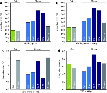
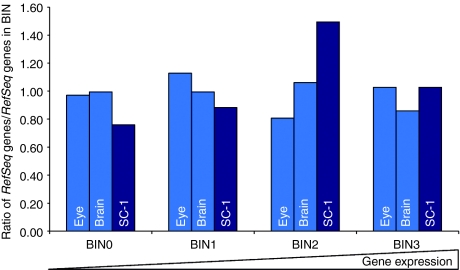
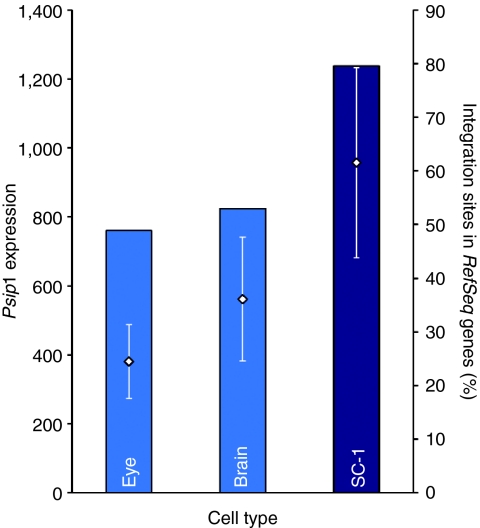
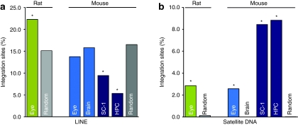
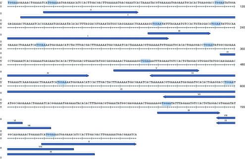
Lentiviral vectors with self-inactivating (SIN) long terminal repeats (LTRs) are promising for safe and sustained transgene expression in dividing as well as quiescent cells. As genome organization and transcription substantially differs between actively dividing and postmitotic cells in vivo, we hypothesized that genomic vector integration preferences might be distinct between these biological states. We performed integration site (IS) analyses on mouse dividing cells (fibroblasts and hematopoietic progenitor cells (HPCs)) transduced ex vivo and postmitotic cells (eye and brain) transduced in vivo. As expected, integration in dividing cells occurred preferably into gene coding regions. In contrast, postmitotic cells showed a close to random frequency of integration into genes and gene spare long interspersed nuclear elements (LINE). Our studies on the potential mechanisms responsible for the detected differences of lentiviral integration suggest that the lowered expression level of Psip1 reduce the integration frequency in vivo into gene coding regions in postmitotic cells. The motif TGGAA might represent one of the factors for preferred lentiviral integration into mouse and rat Satellite DNA. These observations are highly relevant for the correct assessment of preclinical biosafety studies, indicating that lentiviral vectors are well suited for safe and effective clinical gene transfer into postmitotic tissues.


Figures

Figure 1
Figure 1
Lentiviral integration site (IS) distribution in rodent RefSeq genes and the surrounding 10 kbp region. (a) The percentage of lentiviral IS located in RefSeq genes and (b) in RefSeq genes including the surrounding 10 kbp region is shown. IS frequency into RefSeq genes is substantially lower in postmitotic cells compared to dividing cells. Including the 10 kbp vicinity of RefSeq genes, distribution of our postmitotic data sets obtained from retinal cells was not statistically different compared to a random distribution (binomial distribution, two-sided test, P < 0.05). Bars denote frequency of IS retrieved from rat eye (green), from mouse eye and brain tissue (light blue) and from mouse dividing cells (dark blue). The percentages of IS in the vicinity of (c) transcriptional start sites (TSS) and (d) CpG islands are displayed. The distribution of IS in TSS and the 5 kbp surrounding genomic region and in CpG Islands and the surrounding 1 kbp genomic region have been analyzed. Statistical testings have been performed by comparing the experimental data to in silico generated IS, no statistical significant differences compared to the random data set have been observed. The frequency of in silico generated IS located in the respective feature are displayed in light gray for the rat genome and in dark gray for the mouse genome. Of note, to date there are 1.56 times more RefSeq genes known from the mouse genome sequencing project compared to the rat genome. *Statistically different integration site distribution compared to in silico generated random data set (binomial distribution, two-sided test, P < 0.05); kbp, kilobase pair(s); SC-1, mouse fibroblasts; HPC, hematopoietic progenitor cells.
Figure 2
Figure 2
Association between lentiviral integration and gene expression in mouse fibroblasts, mouse eye, and brain tissues. Illumina BeadChip microarray analyses of untransduced dividing and postmitotic mouse cell types revealed a slightly preferred vector integration into (highly) expressed cellular genes for dividing mouse SC-1 fibroblasts (dark blue). The integration preference into expressed genes is less pronounced for mouse postmitotic eyes and brain cells (light blue). The respective Probe IDs with “NM_” accessions on the Illumina MouseRef-8 and Illumina MouseRef-6 Expression BeadChip were grouped and sorted on their expression values (four BINs), (0: likely background expression; 1: low expression; 2: medium expression; 3: medium to high expression). The ratio of integration site within RefSeq genes belonging to one of these four groups to the percentages of overall RefSeq genes which are belonging to these four groups are displayed.
Figure 3
Figure 3
Relation of expression level of Psip1 and integration site (IS) distribution in RefSeq genes. Expression levels of Psip1 were measured on Illumina MouseRef-8 Expression BeadChip microarray on RNA from untransduced mouse SC-1 fibroblasts, postmitotic eye and brain cells, revealing an increased integration frequency into gene coding regions with increasing Psip1 expression. Diamonds display the average signals of Psip1 in the distinct cell types (left y-axis). The percentages of IS located in RefSeq genes, analyzed by pyrosequencing, are shown in bars (right y-axis). Light blue bars are displaying the results obtained from postmitotic tissues, dark blue bars from dividing cells (SC-1). Standard errors of mean signals are indicated as error bars. ProbeID 1780082 is exemplarily shown. SC-1, mouse fibroblasts.
Figure 4
Figure 4
Lentiviral integration site (IS) distribution in rodent repetitive elements. Distinct IS preferences in mouse dividing and postmitotic cells were found for (a) Satellite DNA and (b) LINE. In all data sets, IS distribution in Satellite DNA was significantly overrepresented compared to the random distribution of Satellite DNA in the mouse genome (0.08%). In mouse brain, no IS have been detected in Satellite DNA. IS distribution in LINE corresponded in mouse eye, and in mouse brain tissue to a random distribution (binomial distribution, two-sided test), and was significantly lower for all data sets from dividing cell types compared to in silico generated IS. Two-sided binomial testings for IS obtained from rat eye tissue have been performed on in silico generated IS from the rat genome. A preferred integration in LINE and Satellite DNA has been observed for IS in rat eyes. Green bars denote IS in rat eyes, light blue bars are displaying IS in mouse nondividing cells, and dark blue bars IS in dividing cells. IS in LINE and Satellite DNA generated from an in silico data set are shown in light gray for the rat genome and in dark gray for the mouse genome. HPC, hematopoietic progenitor cells; LINE, long interspersed nuclear element; SC-1, mouse fibroblasts.
Figure 5
Figure 5
Clustering of lentiviral integration sites (IS) in γ-Satellite. The preferred lentiviral integration into mouse (and rat) Satellite DNA observed in dividing and postmitotic cells, except for mouse brain tissue, is exemplarily shown for a prototypical Satellite sequence to demonstrate how close the insertions are to the motif. In this case, identified IS were aligned to a γ-Satellite DNA (GSAT_MM). The genomic part of linear amplification–mediated PCR amplicons which aligned to this Satellite DNA are shown as blue arrows, TGGAA motif are highlighted in light blue. Numbers above the arrows indicate different IS. IS were obtained by Sanger sequencing.

Comment in

References

    1. Zufferey R, Dull T, Mandel RJ, Bukovsky A, Quiroz D, Naldini L.et al. (1998Self-inactivating lentivirus vector for safe and efficient in vivo gene delivery J Virol 729873–9880. - PMC - PubMed
    1. Cartier N, Hacein-Bey-Abina S, Bartholomae CC, Veres G, Schmidt M, Kutschera I.et al. (2009Hematopoietic stem cell gene therapy with a lentiviral vector in X-linked adrenoleukodystrophy Science 326818–823. - PubMed
    1. Montini E, Cesana D, Schmidt M, Sanvito F, Ponzoni M, Bartholomae C.et al. (2006Hematopoietic stem cell gene transfer in a tumor-prone mouse model uncovers low genotoxicity of lentiviral vector integration Nat Biotechnol 24687–696. - PubMed
    1. Montini E, Cesana D, Schmidt M, Sanvito F, Bartholomae CC, Ranzani M.et al. (2009The genotoxic potential of retroviral vectors is strongly modulated by vector design and integration site selection in a mouse model of HSC gene therapy J Clin Invest 119964–975. - PMC - PubMed
    1. Modlich U, Navarro S, Zychlinski D, Maetzig T, Knoess S, Brugman MH.et al. (2009Insertional transformation of hematopoietic cells by self-inactivating lentiviral and gammaretroviral vectors Mol Ther 171919–1928. - PMC - PubMed

Publication types