. 2011 Mar 15;104(6):957-67.
doi: 10.1038/bjc.2011.42. Epub 2011 Mar 1.

Potentiation of apoptosis by histone deacetylase inhibitors and doxorubicin combination: cytoplasmic cathepsin B as a mediator of apoptosis in multiple myeloma

Affiliations

Potentiation of apoptosis by histone deacetylase inhibitors and doxorubicin combination: cytoplasmic cathepsin B as a mediator of apoptosis in multiple myeloma

V Cheriyath et al. Br J Cancer. .

Abstract

Background: Although inhibitors of histone deacetylase inhibitors (HDACis) in combination with genotoxins potentiate apoptosis, the role of proteases other than caspases in this process remained elusive. Therefore, we examined the potentiation of apoptosis and related mechanisms of HDACis and doxorubicin combination in a panel of myeloma cell lines and in 25 primary myelomas.

Results: At IC(50) concentrations, sodium butyrate (an HDACi) or doxorubicin alone caused little apoptosis. However, their combination potentiated apoptosis and synergistically reduced the viability of myeloma cells independent of p53 and caspase 3-7 activation. Potentiated apoptosis correlated with nuclear translocation of apoptosis-inducing factor, suggesting the induction of caspase 3- and 7-independent pathways. Consistent with this, butyrate and doxorubicin combination significantly increased the activity of cytoplasmic cathepsin B. Inhibition of cathepsin B either with a small-molecule inhibitor or downregulation with a siRNA reversed butyrate- and doxorubicin-potentiated apoptosis. Finally, ex vivo, clinically relevant concentrations of butyrate or SAHA (suberoylanilide hydroxamic acid, vorinostat, an HDACi in clinical testing) in combination with doxorubicin significantly (P<0.0001) reduced the survival of primary myeloma cells.

Conclusions: Cathepsin B has a prominent function in mediating apoptosis potentiated by HDACi and doxorubicin combinations in myeloma. Our results support a molecular model of lysosomal-mitochondrial crosstalk in HDACi- and doxorubicin-potentiated apoptosis through the activation of cathepsin B.


Conflict of interest statement

The authors declare no conflict of interest.

Figures

Figure 1
Figure 1
Combinations of HDACi and doxorubicin potentiate apoptosis in myeloma cells. (A) Effects of HDACi, doxorubicin and their combination on apoptosis of myeloma cells with varying p53 status. Myeloma cells (1 × 10⁶) carrying either wild-type or mutant p53 (NCI H929, RPMI 8226, U266, KMS11 and OPM2) were left untreated or treated with butyrate (300 μM for NCI H929 and 600 μM for RPMI 8226, U266, KMS 11 and OPM2) or doxorubicin (40 nM) or with their combination for 48 h. Percentage of cells undergoing apoptosis was assessed by TUNEL staining. Scatter plot shown is a representative of two independent experiments with similar results, in which 10 000 events were collected using flow cytometry. Mutational status of p53 of the myeloma cell lines is indicated. Induction of apoptosis in myeloma cells by butyrate, doxorubicin and their combination are summarised in the bottom graph. Each bar on the graph is mean±s.e.m. of two independent experiments. # Indicates that the treatment is significantly different from other treatments and ‘&’ sign indicates that treatment is significantly different from untreated or butyrate treatment; P-values for each treatment is provided. (B) Effect of butyrate and doxorubicin (40 nM) combination on HDAC activity. Whole-cell lysates (WHL; 30 μg) of NCI-H929 or RPMI 8226 cells left untreated or treated with butyrate (+=300 μM, +*=600 μM) doxorubicin or their combination for 36 h and acetylation status of histone H4 as an indirect measure of HDAC activity was determined by immunoblot analysis.
Figure 2
Figure 2
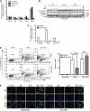
Effects of butyrate, doxorubicin or their combination on caspase 3 and 7 activation and AIF release in myeloma cells. (A, B) Effects of butyrate, doxorubicin and their combination on caspase 3 and 7 activation. (A) NCI H929, RPMI 8226 and U266 cells were treated with butyrate (SB; 300 μM for NCI H929 and 600 μM for RPMI 8226 and U266), doxorubicin (Dox; 40 nM) or with their combination. After 24 h treatments, fold change in caspase 3 and 7 activity relative to untreated cells was assessed by caspase 3 and 7 glo kit (Promega Inc.). TRAIL (50 ng ml−1, Peprotech Inc., Rocky Hill, NJ, USA) was used as a positive control. Each data point in the bar graph is mean±s.e.m. of three independent experiments performed in triplicate. (B) Caspase 3 cleavage was assessed after 16, 24 or 36 h by subjecting 30 μg of whole-cell lysates (WCL) of RPMI 8226 cells to immunoblot analysis with a caspase 3-specific antibody. TRAIL-treated sample was used as a positive control and β-Actin as a loading control. (C, D) Effects of caspase 3 inhibitor DEVD-CHO on butyrate- and doxorubicin-induced apoptosis of myeloma cells. RPMI 8226 cells (1 × 10⁶) were pretreated with either vehicle (DMSO) or 1 μM of cell permeable caspase 3-specific inhibitor DEVD-CHO (Biomol Inc.) for 2 h. Then the cells were left untreated or treated with TRAIL (50 ng ml−1) or butyrate (600 μM) plus doxorubicin (40 nM). Caspase 3 and 7 activity was determined as in Figure 3A, and percentage of cells undergoing apoptosis was determined 48 h post-treatment by TUNEL assay as in Figure 2. Scatter plot shown is one of two independent experiments with similar results, in which 10 000 events were collected (top panel). Each bar on the graph is mean±s.e.m. of two independent experiments, and P-values of significantly different treatments are provided. (E) Butyrate plus doxorubicin combination results in nuclear translocation of AIF in RPMI 8226 and NCI H929 cells. RPMI 8226 or NCI H929 cells were left untreated or treated with indicated concentrations of butyrate, doxorubicin or their combination for 48 h. The localisation of AIF was assessed by indirect immunofluorescence staining with an AIF antibody followed by Alexa Flour-488-conjugated secondary antibody (Green staining). Nuclei of the cells were stained with DAPI (blue). Merged images were produced by superimposing both images. Results shown are representative of three independent experiments with similar results.
Figure 3
Figure 3
Combinations of butyrate and doxorubicin significantly increased the activity of cytoplasmic cathepsin B. (A) Optimisation of cytoplasmic cathepsin B extraction by permeabilisation of plasma membrane with digitonin. RPMI 8226 cells plasma membranes were permeabilised with increasing concentrations of digitonin for 10 min in ice. Permeabilisation of plasma membranes was monitored by assessing LDH activity (left Y axis) and permeabilisation of lysosomes was monitored by cathepsin B activity (right Y axis). Each point on the graph is mean±s.e.m. of two independent experiments. (B) Effects of butyrate and doxorubicin combination on the activity of cytoplasmic cathepsin B in myeloma cells. RPMI 8226 cells were treated with butyrate (SB, 600 μM), doxorubicin (Dox, 40 nM) or their combination. Cells were harvested at indicated time periods, permeabilised with 50 μg ml−1 digitonin, and the activity of cathepsin B was measured using enzyme assay kits (Biovision Inc.). Cathepsin B activity was normalised to LDH activity; *P<0.05, **P<0.001 and ***P<0.0001. (C, D) Effects of butyrate and doxorubicin combination on the activity of calpain and cathepsin D in RPMI 8226 cells. Relative increase in total calpain (C) and cathepsin D (D) activities were calculated by normalising to untreated samples. Each data point on the graph is mean±s.e.m. of two independent experiments performed in triplicate.
Figure 4
Figure 4
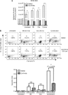
Cathepsin B inhibitor attenuated butyrate- and doxorubicin-induced apoptosis in RPMI 8226 cells. (A) Cathepsin B inhibitor attenuated butyrate- and doxorubicin-mediated activation of cathepsin B in RPMI 8226 cells. RPMI 8226 cells were treated with cell permeable cathepsin B-specific inhibitor CA-074Me (12.5 μM) or vehicle (DMSO) for 1 h before treatment with butyrate (SB; 0.6 mM), doxorubicin (Dox; 40 nM) or combination. After 40 h treatment, cathepsin B activity was assessed using cathepsin B activity assay kit (Biovision Inc). Each data point on the graph is an average of two independent experiments performed in triplicate. (B) Cathepsin B inhibitor antagonised butyrate- and doxorubicin-induced apoptosis. After 72 h butyrate (SB; 0.6 mM), doxorubicin (Dox; 40 nM) or combination treatment, apoptosis indices in vehicle (DMSO)- or CA-074me (12.5 μM)-pretreated cells were compared with TUNEL assay. Scatter plot shown is one of two independent experiments with similar results, in which 10 000 events were collected (top panel). Each bar on the graph is mean±s.e.m. of two independent experiments, and P-value of significantly different treatments is provided (bottom panel). (C, D) Downregulation of cathepsin B with a siRNA rescue RPMI 8226 cells from combination potentiated apoptosis. RPMI 8226 cells were transfected with either a scrambled siRNA (siControl) or a siRNA specific to cathepsin B (siCathepsin B). Knockdown of cathepsin B was determined by immunoblot analysis of 30 μg of WHL 48 h post-transfection (C) and apoptotic index 72 h post-transfection by TUNEL assay (D). Each bar on the graph is mean±s.e.m. of two independent experiments.
Figure 4
Figure 4
Cathepsin B inhibitor attenuated butyrate- and doxorubicin-induced apoptosis in RPMI 8226 cells. (A) Cathepsin B inhibitor attenuated butyrate- and doxorubicin-mediated activation of cathepsin B in RPMI 8226 cells. RPMI 8226 cells were treated with cell permeable cathepsin B-specific inhibitor CA-074Me (12.5 μM) or vehicle (DMSO) for 1 h before treatment with butyrate (SB; 0.6 mM), doxorubicin (Dox; 40 nM) or combination. After 40 h treatment, cathepsin B activity was assessed using cathepsin B activity assay kit (Biovision Inc). Each data point on the graph is an average of two independent experiments performed in triplicate. (B) Cathepsin B inhibitor antagonised butyrate- and doxorubicin-induced apoptosis. After 72 h butyrate (SB; 0.6 mM), doxorubicin (Dox; 40 nM) or combination treatment, apoptosis indices in vehicle (DMSO)- or CA-074me (12.5 μM)-pretreated cells were compared with TUNEL assay. Scatter plot shown is one of two independent experiments with similar results, in which 10 000 events were collected (top panel). Each bar on the graph is mean±s.e.m. of two independent experiments, and P-value of significantly different treatments is provided (bottom panel). (C, D) Downregulation of cathepsin B with a siRNA rescue RPMI 8226 cells from combination potentiated apoptosis. RPMI 8226 cells were transfected with either a scrambled siRNA (siControl) or a siRNA specific to cathepsin B (siCathepsin B). Knockdown of cathepsin B was determined by immunoblot analysis of 30 μg of WHL 48 h post-transfection (C) and apoptotic index 72 h post-transfection by TUNEL assay (D). Each bar on the graph is mean±s.e.m. of two independent experiments.

Similar articles

Cited by

References

    1. Badros A, Burger AM, Philip S, Niesvizky R, Kolla SS, Goloubeva O, Harris C, Zwiebel J, Wright JJ, Espinoza-Delgado I, Baer MR, Holleran JL, Egorin MJ, Grant S (2009) Phase I study of vorinostat in combination with bortezomib for relapsed and refractory multiple myeloma. Clin Cancer Res 15(16): 5250–5257 - PMC - PubMed
    1. Bae SI, Cheriyath V, Jacobs BS, Reu FJ, Borden EC (2008) Reversal of methylation silencing of Apo2L/TRAIL receptor 1 (DR4) expression overcomes resistance of SK-MEL-3 and SK-MEL-28 melanoma cells to interferons (IFNs) or Apo2L/TRAIL. Oncogene 27(4): 490–498 - PubMed
    1. Balliet RM, Chen G, Gallagher CJ, Dellinger RW, Sun D, Lazarus P (2009) Characterization of UGTs active against SAHA and association between SAHA glucuronidation activity phenotype with UGT genotype. Cancer Res 69(7): 2981–2989 - PMC - PubMed
    1. Bergsagel PL, Kuehl WM (2001) Chromosome translocations in multiple myeloma. Oncogene 20(40): 5611–5622 - PubMed
    1. Bidere N, Lorenzo HK, Carmona S, Laforge M, Harper F, Dumont C, Senik A (2003) Cathepsin D triggers Bax activation, resulting in selective apoptosis-inducing factor (AIF) relocation in T lymphocytes entering the early commitment phase to apoptosis. J Biol Chem 278(33): 31401–31411 - PubMed

Publication types

MeSH terms