Review
. 2010;1(1):e9.
doi: 10.1038/cddis.2009.11.

Long-term (trophic) purinergic signalling: purinoceptors control cell proliferation, differentiation and death

Affiliations
Review

Long-term (trophic) purinergic signalling: purinoceptors control cell proliferation, differentiation and death

G Burnstock et al. Cell Death Dis. 2010.

Abstract

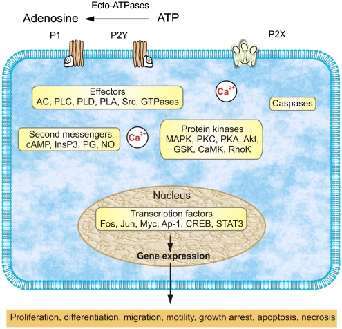
The purinergic signalling system, which uses purines and pyrimidines as chemical transmitters, and purinoceptors as effectors, is deeply rooted in evolution and development and is a pivotal factor in cell communication. The ATP and its derivatives function as a 'danger signal' in the most primitive forms of life. Purinoceptors are extraordinarily widely distributed in all cell types and tissues and they are involved in the regulation of an even more extraordinary number of biological processes. In addition to fast purinergic signalling in neurotransmission, neuromodulation and secretion, there is long-term (trophic) purinergic signalling involving cell proliferation, differentiation, motility and death in the development and regeneration of most systems of the body. In this article, we focus on the latter in the immune/defence system, in stratified epithelia in visceral organs and skin, embryological development, bone formation and resorption, as well as in cancer.


Figures

Figure 1
Figure 1
Overview of purinergic signalling mechanisms that regulate long-term, trophic effects. Extracellular nucleotides and nucleosides bind to purinoceptors coupled to signal-transducing effector molecules. Activation of effectors leads to generation of second messengers and/or stimulation of protein kinases that regulate expression of genes needed for long-term, trophic actions. Trophic action of P2X receptors can be mediated by increases in cytosolic Ca2+ concentration; activation of P2X7 receptors can also be coupled to protein kinase cascades and caspases that can mediate proliferation and apoptosis. Cell-specific and/or receptor subtype-specific differences are likely to account for variations in signalling pathways and functional outcomes. It should be noted that the list of elements is not meant to be all-inclusive. Other protein kinases, for example, MEK and PI3K, are upstream of the listed kinases involved in purinergic signalling, whereas others are downstream, for example, p70S6K. In addition, dashed arrows indicate that not all listed elements are activated by the upstream component, for example, not all P1 receptors are coupled to all listed effectors. AC, adenylyl cyclase; AP-1, activator protein-1; CaMK, calcium/calmodulin protein kinase; CREB, cAMP response element binding protein; DG, diacylglycerol; GSK, glycogen synthase kinase; InsP3, inositol trisphosphate; MAPKs, mitogen-activated protein kinases (including extracellular signal-regulated protein kinase (ERK), p38 MAPK, and stress-activated protein kinase (SAPK)/c-Jun NH2-terminal kinase (JNK)); MEK, MAPK/ERK kinase; NO, nitric oxide; PG, prostaglandin; PI3K, phosphoinositide 3-kinase; PLC, phosphatidylinositol-specific phospholipase C; PKA, protein kinase A; PKC, protein kinase C; PLD, phospholipase D; PLA, phospholipase A; STAT3, signal transducer and activator of transcription-3 (based on Figure 11 from Burnstock with permission from the American Physiological Society )
Figure 2
Figure 2
Double labelling of P2Y1 and P2Y2 receptors with markers of proliferation shows colocalisation within a sub-population of basal and parabasal keratinocytes. Double labelling of P2X5 receptors with markers of differentiated keratinocytes shows colocalisation within the stratum spinosum, and double labelling of P2X7 receptors with markers of apoptosis in human leg skin shows colocalisation within the stratum corneum. (a) Ki-67 immunolabelling (a marker for proliferation) stained the nuclei (green) of a sub-population of keratinocytes in the basal and parabasal layers of the epidermis. P2Y1 receptor immunostaining (red) was found in the basal layer on cells also staining for Ki67. Scale bar 30 μm. (b) PCNA immunolabelling (a marker for proliferation) stained the nuclei (green) of a sub-population of keratinocytes. These nuclei were often distributed in clusters and found in the basal and parabasal layers of the epidermis. P2Y2 receptor immunostaining (red) was also expressed in basal and parabasal epidermal cells. Scale bar 30 μm. (c) P2X5 receptor immunostaining (red) showed overlap (yellow) with cytokeratin K10 (green), an early marker of keratinocyte differentiation. P2X5 receptors were present in the basal layer of the epidermis up to the mid-granular layer. Cytokeratin K10 was distributed in most suprabasal keratinocytes. The stratum basale stained only for P2X5 receptors, indicating that no differentiation was taking place in these cells. The colocalisation of P2X5 receptors and cytokeratin K10 appeared mainly in the cytoplasm of differentiating cells within the stratum spinosum and partly in the stratum granulosum. Note that the stratum corneum also stained for cytokeratin K10, which labelled differentiated keratinocytes, even in dying cells. Scale bar 30 μm. (d) P2X5 receptor immunostaining (red) showed overlap (yellow) with involucrin (green). P2X5 receptors were present in the basal layer of the epidermis up to the mid-granular layer. Note that the pattern of staining with involucrin was similar to that seen with cytokeratin K10, except that cells from the stratum basale up to the midstratum spinosum were not labelled with involucrin, which is a late marker of keratinocyte differentiation. Scale bar 30 μm. (e) TUNEL (green) labelled the nuclei of cells at the uppermost level of the stratum granulosum and P2X7 antibody (red) mainly stained cell fragments within the stratum corneum. Scale bar 15 μm. (f) Anti-caspase-3 (green) colocalised with areas of P2X7 receptor immunostaining (red) both at the junction of the stratum granulosum and within the stratum corneum. Areas of colocalisation were yellow. Note that the differentiating keratinocytes in the upper stratum granulosum were also positive for anti-caspase-3. Scale bar 15 μm (reproduced with permission from Greig et al.)
Figure 3
Figure 3
At 48 h after application of drugs to primary human keratinocyte cultures. (a) ATP (1–10 μM) and UTP (100 μM) cause an increase in cell number, whereas ATPγS (100–500 μM) and ATP (100 μM) cause a significant decrease. Results represent the mean of eight experiments. *P<0.001 compared with that of control. (b) 2MeSADP (500 μM) causes a significant increase in cell number. Results represent the mean of eight experiments. *P<0.05 compared with that of control. (c) BzATP (100–500 μM) causes a significant decrease in cell number. Results represent the mean of nine experiments. *P<0.001 compared with that of control. Error bars represent mean±S.E.M (reproduced with permission from Greig et al.)
Figure 4
Figure 4
Schematic diagram illustrating the different mechanisms by which P2 receptor subtypes might alter cancer cell function. P2Y1 and P2Y2 receptors could affect the rate of cell proliferation through altering the intracellular levels of cAMP by modulating adenylyl cyclase (AC) or by increasing intracellular calcium levels through the phospholipase C (PLC) pathway. P2X5 and P2Y11 receptor activation might switch the cell cycle from proliferation into a state of differentiation. The P2X7 receptor activates the apoptotic caspase enzyme system (redrawn from White and Burnstock with permission from Elsevier)
Figure 5
Figure 5
Schematic diagram illustrating the potential functions of extracellular nucleotides and P2 receptors in modulating bone cell function. ATP released from osteoclasts (e.g., through shear stress or constitutively) or from other sources, can be degraded to adenosine 5′-diphosphate (ADP) or converted into uridine 5′-triphosphate (UTP) through ecto-nucleotidases. All three nucleotides can function separately on specific P2 receptor subtypes, as indicated by the colour coding. ATP is a universal agonist, whereas UTP is only active at the P2Y2 receptor and ADP is only active at the P2Y1 receptor. ADP acting on P2Y1 receptors seems to stimulate both the formation (i.e., fusion) of osteoclasts from haematopoietic precursors and the resorptive activity of mature osteoclasts. For the latter, a synergistic action of ATP and protons has been proposed by the P2X2 receptor. ADP could also stimulate resorption indirectly through actions on osteoclasts, which in turn release pro-resorptive factors (e.g., receptor activator of nuclear factor κB ligand, RANKL) ATP at high concentrations might facilitate fusion of osteoclast progenitors through P2X7 receptor pore formation or induce cell death of mature osteoclasts through P2X7 receptors. In osteoblasts, ATP, through P2X5 receptors, might enhance proliferation and/or differentiation. By contrast, UTP, through P2Y2 receptors, is a strong inhibitor of bone formation by osteoblasts. For some receptors (e.g., P2X4 and P2Y2 receptors on osteoclasts or P2X2 receptors on osteoblasts), evidence for expression has been found but their role is still unclear (based on schemes from Hoebertz et al.)

References

    1. Lohmann K. Uber die Pyrophosphatfraktion im Muskel. Naturwissenschaften. 1929;17:624–625.
    1. Fiske CH, SubbaRow Y. Phosphorous compounds of muscle and liver. Science. 1929;70:381–382. - PubMed
    1. Lippman F. Metabolic generation and utilization of phosphate bond energy. Enzymology. 1941;1:99.
    1. Ponnamperuma C, Sagan C, Mariner R. Synthesis of adenosine triphosphate under possible primitive earth conditions. Nature. 1963;199:222–226. - PubMed
    1. Waldrop MM. Did life really start out in an RNA world. Science. 1989;246:1248–1249. - PubMed

Substances