. 2010 May 6;1(5):e40.
doi: 10.1038/cddis.2010.18.

A survey of the anti-apoptotic Bcl-2 subfamily expression in cancer types provides a platform to predict the efficacy of Bcl-2 antagonists in cancer therapy

Affiliations

A survey of the anti-apoptotic Bcl-2 subfamily expression in cancer types provides a platform to predict the efficacy of Bcl-2 antagonists in cancer therapy

W J Placzek et al. Cell Death Dis. .

Abstract

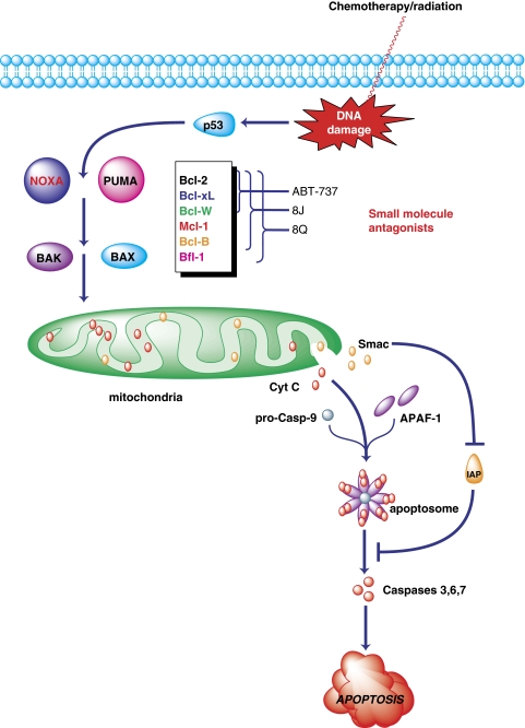
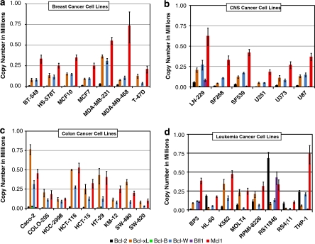
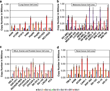
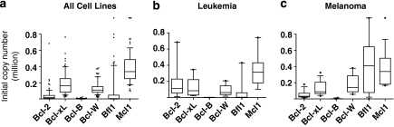
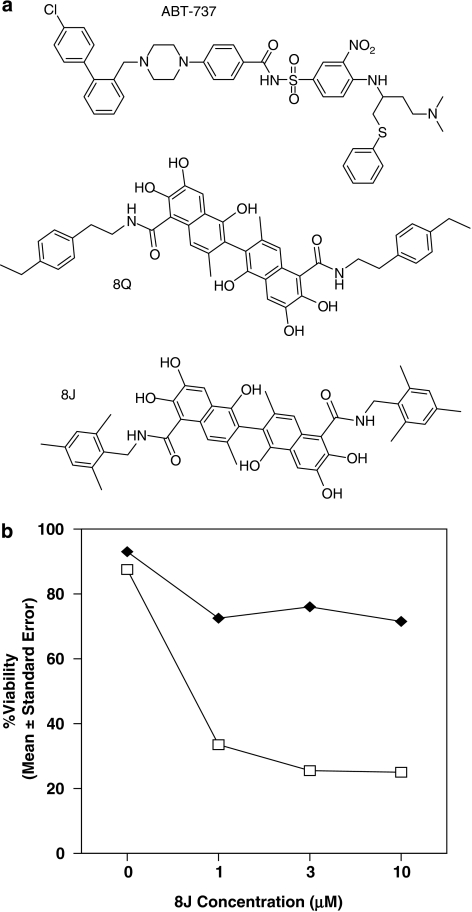
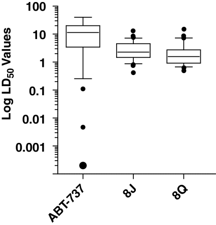
We investigated the mRNA expression levels of all six antiapoptotic Bcl-2 subfamily members in 68 human cancer cell lines using qPCR techniques and measured the ability of known Bcl-2 inhibitors to induce cell death in 36 of the studied tumor cell lines. Our study reveals that Mcl-1 represents the anti-apoptotic Bcl-2 subfamily member with the highest mRNA levels in the lung, prostate, breast, ovarian, renal, and glioma cancer cell lines. In leukemia/lymphoma and melanoma cancer cell lines, Bcl-2 and Bfl-1 had the highest levels of mRNA, respectively. The observed correlation between the cell killing properties of known Bcl-2 inhibitors and the relative mRNA expression levels of anti-apoptotic Bcl-2 proteins provide critical insights into apoptosis-based anticancer strategies that target Bcl-2 proteins. Our data may explain current challenges of selective Bcl-2 inhibitors in the clinic, given that severe expression of Bcl-2 seems to be limited to leukemia cell lines. Furthermore, our data suggest that in most cancer types a strategy targeted to Mcl-1 inhibition, or combination of Bfl-1 and Mcl-1 inhibition for melanoma, may prove to be more successful than therapies targeting only Bcl-2.


Figures

Figure 1
Figure 1
Mechanism of Bcl-2 antagonists in cell stress response. The overexpression of antiapoptotic Bcl-2 proteins induce cancer cell resistance to stress-induced apoptosis. Selective inhibition of a subset of the antiapoptotic Bcl-2 subfamily, as is achieved with ABT-737, has provided promising clinical outcomes, but has also been shown to be overcome through upregulation of Mcl-1 or Bfl-1. Pan-active inhibition of all members of the antiapoptotic Bcl-2 subfamily, as is achieved by the Apogossypol derivative, 8Q, prevents suppression of p53-mediated apoptosis and precludes compound resistance through upregulation of other subfamily members
Figure 2
Figure 2
Expression of the six antiapoptotic Bcl-2 subfamily members in the breast, CNS, colon, and leukemia cancer cell lines. (ad) Bar graphs representing the relative copy number of each Bcl-2 subfamily member: Bcl-2 in black, Bcl-xL in orange, Bcl-B in green, Bcl-W in blue, Bfl1 in purple, and Mcl1 in red. Cell lines are grouped according to their identified tissue type. Error bars represent S.E.M.
Figure 3
Figure 3
Expression of the six antiapoptotic Bcl-2 subfamily members in the lung, melanoma, ovarian, prostate, and renal cancer cell lines. (ad) Bar graphs representing the relative copy number of each Bcl-2 subfamily member: Bcl-2 in black, Bcl-xL in orange, Bcl-B in green, Bcl-W in blue, Bfl1 in purple, and Mcl1 in red. Cell lines are grouped according to their identified tissue type. Error bars represent S.E.M.
Figure 4
Figure 4
Range of antiapoptotic Bcl-2 subfamily expression. (ac) Box and Whisker plot of the 10–90th percentile of the relative copy number distribution across all 68 cell lines studies (a), the 8 leukemia cell lines (b), or the 12 melanoma cell lines (c)
Figure 5
Figure 5
Chemical structures of Bcl-2 antagonists. (a) Chemical structures of the three Bcl-2 inhibitors studied. (b) Cell viability of WT-MEF cells (white squares) or bax−/−bak−/− DKO MEF cells (solid squares) treated with compound 8J at varying concentrations
Figure 6
Figure 6
Ability of cells to evade apoptosis in presence of Bcl-2 inhibitors. (a) LD50 values for each of the three compounds ABT-737 (blue bars), 8J (red bars), and 8Q (green bars) in 36 selected cell lines grouped according to their cancer type are shown. Error bars represent S.E.M. and the asterisks represent cell lines in which ABT-737 showed no apparent cell killing effect at a concentration of 30 μM. (b) Scatter plot of Bcl-2 mRNA copy number and the measured LD50 values for the 36 cell selected cell lines
Figure 7
Figure 7
Range of compound LD50 values across the 36 cell lines studied. Box and Whisker plot of the 10–90th percentile of the measured LD50 values for the three compounds are represented on a log scale

References

    1. Reed JC. Dysregulation of apoptosis in cancer. J Clin Oncol. 1999;17:2941–2953. - PubMed
    1. Adams JM, Cory S. The Bcl-2 protein family: arbiters of cell survival. Science. 1998;281:1322–1326. - PubMed
    1. Liang H, Fesik SW. Three-dimensional structures of proteins involved in programmed cell death. J Mol Biol. 1997;274:291–302. - PubMed
    1. Green DR, Kroemer G. The pathophysiology of mitochondrial cell death. Science. 2004;305:626–629. - PubMed
    1. Fesik SW. Promoting apoptosis as a strategy for cancer drug discovery. Nat Rev Cancer. 2005;5:876–885. - PubMed

Publication types

MeSH terms

Substances