. 2010 Jul 1;1(7):e51.
doi: 10.1038/cddis.2010.25.

An otoprotective role for the apoptosis inhibitor protein survivin

Affiliations

An otoprotective role for the apoptosis inhibitor protein survivin

S K Knauer et al. Cell Death Dis. .

Abstract

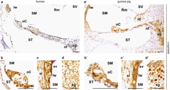
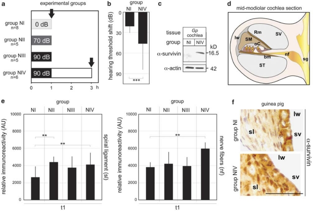
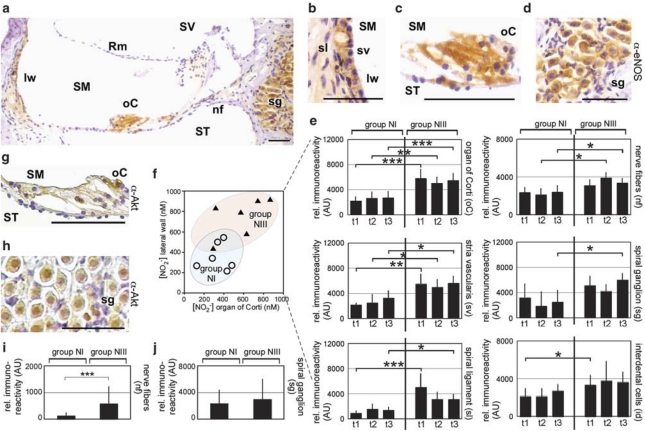
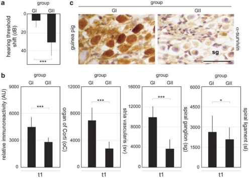
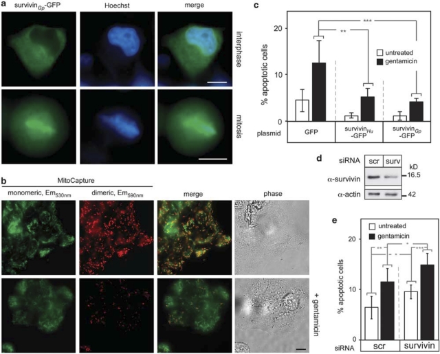
Hearing impairment caused by ototoxic insults, such as noise or gentamicin is a worldwide health problem. As the molecular circuitries involved are not yet resolved, current otoprotective therapies are rather empirical than rational. Here, immunohistochemistry and western blotting showed that the cytoprotective protein survivin is expressed in the human and guinea pig cochlea. In the guinea pig model, moderate noise exposure causing only a temporary hearing impairment transiently evoked survivin expression in the spiral ligament, nerve fibers and the organ of Corti. Mechanistically, survivin upregulation may involve nitric oxide (NO)-induced Akt signaling, as enhanced expression of the endothelial NO synthase and phosphorylated Akt were detectable in some surviving-positive cell types. In contrast, intratympanic gentamicin injection inducing cell damage and permanent hearing loss correlated with attenuated survivin levels in the cochlea. Subsequently, the protective activity of the human and the guinea pig survivin orthologs against the ototoxin gentamicin was demonstrated by ectopic overexpression and RNAi-mediated depletion studies in auditory cells in vitro. These data suggest that survivin represents an innate cytoprotective resistor against stress conditions in the auditory system. The pharmacogenetic modulation of survivin may thus provide the conceptual basis for the rational design of novel therapeutic otoprotective strategies.


Figures

Figure 1
Figure 1
Survivin expression in various regions of the human (ad) and the guinea pig (a′d′) cochlea visualized by IHC. (a/a′) Overview. (b/b′) Survivin is detectable in all cell types in the organ of Corti, except the cuticular plates of pillar cells and in Claudius cells. (c/c′) In the lateral wall, survivin immunoreactivity was observed in the spiral ligament and the stria vascularis. (d/d′) Survivin expression in the spiral ganglions. id, interdental cells; is, inner sulcus; lw, lateral wall; nf, nerve fibers; oC, organ of Corti; os, outer sulcus; Rm, Reissner's membrane; sg, spiral ganglion; sl, spiral ligament; SM, scala media; ST, scala tympany; sv, stria vascularis; SV, scala vestibuli. Scale bars, 50 μm
Figure 2
Figure 2
Survivin induction by noise exposure. (a) Overview of treatment and animal groups. Guinea pigs were either unexposed (NI) or exposed to 70 dB (NII) or 90 dB (NIII/IV) SPL for 1 h. Bullae were removed at the indicated time points (arrows). (b) ABR recording revealed a mean hearing impairment of 33 dB in animals 2 h after noise exposure (90 dB SPL for 1 h, n=6, group NVI) compared to unexposed controls (n=8, group NI). Columns, mean; bars, ±S.D. ***P<0.005. (c) Noise-induced survivin expression in whole cochleae lysates shown by immunoblot analysis using the α-survivin Ab. Actin served as loading control. (d) Schematics of a cochlear mid-modiolar section. The cochlea is divided into three fluid compartments: the scala vestibuli (SV), the scala media (SM) and the scala tympany (ST), seperated by the Reissner's (Rm) and the basal membrane (bm), respectively. The outer wall of the SM is lined by the lateral wall (lw). The organ of Corti (oC) contains the sensory epithelia comprised of the auditory hair cells, which are innervated by the nerve fibers (nf), and the spiral ganglion neurons (sg). (e/f) Survivin expression before and after acoustic trauma analyzed by IHC. Relative immunoreactivity is indicated for the first cochlea turn (t1). (e) Enhanced survivin levels were evident in the spiral ligament as well as in the nerve fibers already 1 h after acoustic trauma, and further increased 2 h after 90 dB SPL exposure. Columns, mean; bars, ±S.D. **P<0.01. (f) Representative IHC-micrograph demonstrating enhanced survivin levels in the spiral ligament following acoustic trauma. Scale bar, 50 μm
Figure 3
Figure 3
eNOS and NO levels are enhanced by acoustic trauma. Columns, mean; bars, ±S.D. *P<0.05; **P<0.01; ***P<0.005. (ae) eNOS expression in the guinea pig cochlea visualized by IHC. (a) Overview. (bd) eNOS was detectable in all cell types in the stria vascularis, the organ of Corti, nerve fibers and spiral ganglion. Scale bars, 50 μm. (e) eNOS expression before (NI) and after noise (NIII) in the indicated cell types analyzed by IHC. Relative immunoreactivity is indicated for the different cochlea turns. (f) Enhanced [NO2] measured in the supernatant of organ cultures from noise exposed (NIII) versus control animals (NI). (g, h) p-Akt expression visualized by IHC in the organ of Corti, nerve fibers and spiral ganglion following noise exposure (1 h, 90 db SPL). Scale bars, 50 μm. (i, j) eNOS expression before (NI) and after noise (NIII) in the indicated cell types for the first cochlea turn
Figure 4
Figure 4
Gentamicin treatment results in survivin downregulation. Columns, mean; bars, ±S.D. *P<0.05, ***P<0.005. (a) ABR recording revealed a permanent mean hearing impairment of 24 dB in gentamicin (group GI, n=7) versus saline-injected control animals (group GII, n=9) 7 days post treatment. (b) Survivin levels in GI and GII were quantitated by IHC for the first cochlea turn 7 day post injection. (c) Representative IHC micrograph illustrating reduced survivin expression in ganglion cells following gentamicin treatment. Scale bar, 50 μm
Figure 5
Figure 5
Survivin protects against ototoxin-induced cytotoxicity. Columns, mean; bars, ±S.D. from three independent experiments. *P<0.05, **P<0.01, ***P<0.005. (a) Localization of guinea pig survivinGp-GFP transfectants in interphase and mitotic HeLa cells analyzed by fluorescence microscopy. DNA was marked by Hoechst dye (blue). Scale bars, 10 μm. (b) Induction of apoptosis by gentamicin treatment. In untreated HEI-OC1 cells, mitochondrial integrity is shown by the presence of the mono- and dimeric form of the MitoCapture dye (upper panel). Gentamicin treatment (1 μM, 24 h) causes loss of mitochondrial integrity resulting in loss of dimeric MitoCapture dye staining (lower panel). Scale bar, 10 μm. (c) Gentamicin-induced PCD is reduced in survivinGp- and survivinHu-GFP expressing transfectants. Cells transfected with the indicated plasmids were treated with gentamicin (1 μM), and analyzed 24 h later. Fragmented nuclei were visualized by staining with Hoechst dye and the percentage of apoptotic cells counted in at least 500 cells. (d) RNAi-mediated ablation of survivin following transfection with survivin- (surv) or a scrambled control-siRNA (scr) confirmed by immunoblot analysis. Actin served as loading control. (e) RNAi-mediated attenuation of survivin enhanced gentamicin-induced apoptosis. Cells were transfected with the indicated siRNAs and treated with gentamicin (1 μM, for 24 h) 24 h later. Fragmented nuclei were visualized by staining with Hoechst dye, and the percentage of apoptotic cells counted in at least 500 cells

References

    1. Guthrie OW. Aminoglycoside induced ototoxicity. Toxicology. 2008;249:91–96. - PubMed
    1. Eisen MD, Ryugo DK. Hearing molecules: contributions from genetic deafness. Cell Mol Life Sci. 2007;64:566–580. - PMC - PubMed
    1. Sha SH, Qiu JH, Schacht J. Aspirin to prevent gentamicin-induced hearing loss. N Engl J Med. 2006;354:1856–1857. - PubMed
    1. Henderson D, Bielefeld EC, Harris KC, Hu BH. The role of oxidative stress in noise-induced hearing loss. Ear Hear. 2006;27:1–19. - PubMed
    1. Jiang H, Sha SH, Forge A, Schacht J. Caspase-independent pathways of hair cell death induced by kanamycin in vivo. Cell Death Differ. 2006;13:20–30. - PMC - PubMed

Publication types

MeSH terms