. 2010 Jul 8;1(7):e52.
doi: 10.1038/cddis.2010.29.

Endoplasmic reticulum stress inhibition protects steatotic and non-steatotic livers in partial hepatectomy under ischemia-reperfusion

Affiliations

Endoplasmic reticulum stress inhibition protects steatotic and non-steatotic livers in partial hepatectomy under ischemia-reperfusion

I Ben Mosbah et al. Cell Death Dis. .

Abstract

During partial hepatectomy, ischemia-reperfusion (I/R) is commonly applied in clinical practice to reduce blood flow. Steatotic livers show impaired regenerative response and reduced tolerance to hepatic injury. We examined the effects of tauroursodeoxycholic acid (TUDCA) and 4-phenyl butyric acid (PBA) in steatotic and non-steatotic livers during partial hepatectomy under I/R (PH+I/R). Their effects on the induction of unfolded protein response (UPR) and endoplasmic reticulum (ER) stress were also evaluated. We report that PBA, and especially TUDCA, reduced inflammation, apoptosis and necrosis, and improved liver regeneration in both liver types. Both compounds, especially TUDCA, protected both liver types against ER damage, as they reduced the activation of two of the three pathways of UPR (namely inositol-requiring enzyme and PKR-like ER kinase) and their target molecules caspase 12, c-Jun N-terminal kinase and C/EBP homologous protein-10. Only TUDCA, possibly mediated by extracellular signal-regulated kinase upregulation, inactivated glycogen synthase kinase-3β. This is turn, inactivated mitochondrial voltage-dependent anion channel, reduced cytochrome c release from the mitochondria and caspase 9 activation and protected both liver types against mitochondrial damage. These findings indicate that chemical chaperones, especially TUDCA, could protect steatotic and non-steatotic livers against injury and regeneration failure after PH+I/R.


Figures
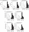
Figure 1
Figure 1
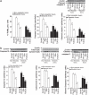
Hepatic damage and inflammatory response. (a) Transaminases and damage score, (b) TNFα and IL1β levels and (c) MPO and MDA levels were measured in both liver types. *P<0.05 versus sham; +P<0.05 versus PH+I/R
Figure 2
Figure 2
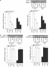
Apoptosis cell death, endoplasmic reticulum and mitochondrial apoptotic cascade. (a) Percentage of TUNEL-positive hepatocytes, the expression of caspase 3 activity and cleaved caspase 3 in both liver types. (b) Expression of caspase 12, cytochrome c and cleaved caspase 9 in both liver types. Representative western blots of cleaved caspase 3, caspase 12, cytochrome c and cleaved caspase 9 at the top and densitometric analysis at the bottom. *P<0.05 versus sham; +P<0.05 versus PH+I/R
Figure 3
Figure 3
Endoplasmic reticulum and mitochondrial damage. (a) Ultrastructural injury score of the endoplasmic reticulum and mitochondria analyzed in both liver types. *P<0.05 versus SHAM; +P<0.05 versus PH+I/R (b) Electron microscopic images of steatotic livers. PH+I/R group: evident ultrastructural alterations of RE and the mitochondria. PBA group: minimal structural alterations of RE and mitochondrial damage similar to the PH+I/R group; TUDCA group: no apparent ultrastructural alterations of RE and the mitochondria
Figure 4
Figure 4
Liver regeneration. The percentage of PCNA-positive hepatocytes was calculated, and HGF and active TGFβ levels were measured in both liver types. *P<0.05 versus sham; +P<0.05 versus PH+I/R
Figure 5
Figure 5
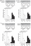
Induction of GRP78 and ATF6 in both liver types. (a) GRP78 mRNA expression and GRP78 protein levels were analyzed in both liver types. For GRP78 mRNA expression in the liver, PCR fluorescent signals for GRP78 were standardized to PCR fluorescent signals obtained from an endogenous reference (β-actin). Comparative and relative quantifications of GRP78 gene products normalized to β-actin and control sham group were calculated by the 2−ΔΔCT method. (b) p50ATF6α and p50ATF6β protein levels were analyzed in both liver types. For GRP78 and ATF6 protein levels in the liver, representative western blots at the top and densitometric analysis at the bottom. *P<0.05 versus sham; +P<0.05 versus PH+I/R. #P<0.05 versus PH+I/R non-steatotic group. GRP78 mRNA and protein levels are significantly lower in the untreated steatotic livers group than in the untreated non-steatotic liver group (#P<0.05)
Figure 6
Figure 6
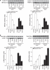
IRE1 pathway. Protein levels of (a) XBP-1 and TRAF2 and (b) pJNK and pP38 were analyzed in both liver types. Representative western blots at the top and densitometric analysis at the bottom. *P<0.05 versus sham; +P<0.05 versus PH+I/R; #P<0.05 versus PH+I/R non-steatotic group. Protein levels of XBP-1 and TRAF2 are significantly lower in the untreated steatotic livers group than in the untreated non-steatotic liver group (#P<0.05)
Figure 7
Figure 7
PERK pathway. Protein levels of (a) pPERK and peIF2α and (b) ATF4 and CHOP in both liver types. Representative western blots at the top and densitometric analysis at the bottom. *P<0.05 versus sham; +P<0.05 versus PH+I/R; #P<0.05 versus PH+I/R non-steatotic group protein levels of PERK, eIF2α, ATF4 and CHOP are significantly lower in the untreated steatotic livers group than in the untreated non-steatotic liver group (#P<0.05)
Figure 8
Figure 8
Endoplasmic reticulum stress and mitochondrial damage. Protein levels of (a) pGSK3β and pVDAC and (b) pERK in both liver types. Representative western blots at the top and densitometric analysis at the bottom. *P<0.05 versus sham; +P<0.05 versus PH+I/R

References

    1. Ramalho FS, Alfany-Fernandez I, Casillas-Ramirez A, Massip-Salcedo M, Serafin A, Rimola A, et al. Are angiotensin II receptor antagonists useful strategies in steatotic and nonsteatotic livers in conditions of partial hepatectomy under ischemia-reperfusion. J Pharmacol Exp Ther. 2009;329:130–140. - PubMed
    1. Kammoun HL, Chabanon H, Hainault I, Luquet S, Magnan C, Koike T, et al. GRP78 expression inhibits insulin and ER stress-induced SREBP-1c activation and reduces hepatic steatosis in mice. J Clin Invest. 2009;119:1201–1215. - PMC - PubMed
    1. Xu C, Bailly-Maitre B, Reed JC. Endoplasmic reticulum stress: cell life and death decisions. J Clin Invest. 2005;115:2656–2664. - PMC - PubMed
    1. Ozcan U, Yilmaz E, Ozcan L, Furuhashi M, Vaillancourt E, Smith RO, et al. Chemical chaperones reduce ER stress and restore glucose homeostasis in a mouse model of type 2 diabetes. Science. 2006;313:1137–1140. - PMC - PubMed
    1. Yoneda T, Imaizumi K, Oono K, Yui D, Gomi F, Katayama T, et al. Activation of caspase-12, an endoplastic reticulum (ER) resident caspase, through tumor necrosis factor receptor-associated factor 2-dependent mechanism in response to the ER stress. J Biol Chem. 2001;276:13935–13940. - PubMed

Publication types

MeSH terms