. 2010 Jul 29;1(7):e60.
doi: 10.1038/cddis.2010.37.

MEK-ERK-dependent multiple caspase activation by mitochondrial proapoptotic Bcl-2 family proteins is essential for heavy ion irradiation-induced glioma cell death

Affiliations

MEK-ERK-dependent multiple caspase activation by mitochondrial proapoptotic Bcl-2 family proteins is essential for heavy ion irradiation-induced glioma cell death

A Tomiyama et al. Cell Death Dis. .

Abstract

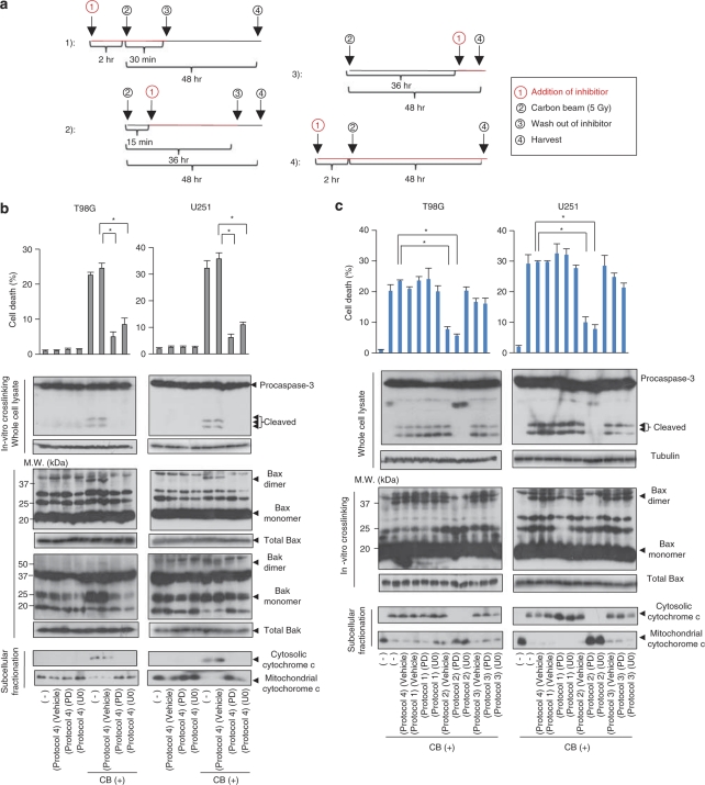
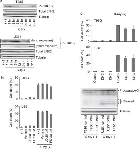
Recently developed heavy ion irradiation therapy using a carbon beam (CB) against systemic malignancy has numerous advantages. However, the clinical results of CB therapy against glioblastoma still have room for improvement. Therefore, we tried to clarify the molecular mechanism of CB-induced glioma cell death. T98G and U251 human glioblastoma cell lines were irradiated by CB, and caspase-dependent apoptosis was induced in both cell lines in a dose-dependent manner. Knockdown of Bax (BCL-2-associated X protein) and Bak (BCL-2-associated killer) and overexpression of Bcl-2 or Bcl-xl (B-cell lymphoma-extra large) showed the involvement of Bcl-2 family proteins upstream of caspase activation, including caspase-8, in CB-induced glioma cell death. We also detected the activation of extracellular signal-regulated kinase (ERK) and the knockdown of ERK regulator mitogen-activated protein kinase kinase (MEK)1/2 or overexpression of a dominant-negative (DN) ERK inhibited CB-induced glioma cell death upstream of the mitochondria. In addition, application of MEK-specific inhibitors for defined periods showed that the recovery of activation of ERK between 2 and 36 h after irradiation is essential for CB-induced glioma cell death. Furthermore, MEK inhibitors or overexpression of a DN ERK failed to significantly inhibit X-ray-induced T98G and U251 cell death. These results suggested that the MEK-ERK cascade has a crucial role in CB-induced glioma cell death, which is known to have a limited contribution to X-ray-induced glioma cell death.


Figures

Figure 1
Figure 1
CB irradiation induces multiple caspase-dependent apoptosis in T98G and U251 glioma cells. (a) T98G and U251 cells treated with or without CB irradiation at the indicated doses were stained with Hoechst 33342 or propidium iodide (PI), and the percentages of dead cells and apoptotic/dead cells were counted using a microscope, as described in the ‘Materials and methods' section at 48 h after irradiation. (b) T98G and U251 cells were CB irradiated (5 Gy) and harvested at the indicated time points after irradiation. The whole-cell lysates obtained were subjected to immunoblotting using the indicated antibodies. As a control of irradiation and DNA damage-induced responses, total cell lysates of T98G and U251 cells obtained at the indicated time periods after X-ray irradiation (20 Gy) or UVC (UV, 200 J) were also subjected to immunoblotting using the same antibodies. The membranes were reprobed using an α-tubulin antibody to confirm equal protein loading. For the detection of cytochrome c release from the mitochondria into the cytosol, T98G and U251 cells were treated by the same stimuli, and then cell lysates obtained at the indicated time points were fractionated into cytosol- and mitochondria-rich fractions as described in the ‘Materials and methods' section and were subjected to immunoblotting using an anti-cytochrome c antibody. To check for equal protein loading, the membranes were reprobed using organelle-specific antibodies (anti-α-tubulin for the cytosol and anti-HSP60 for the mitochondria). (c) T98G and U251 cells were CB irradiated (5 Gy) and harvested at the indicated time points after irradiation. The total cell lysates obtained were subjected to immunoblotting using anti-caspase-8 and anti-Bid antibodies. As a control for extrinsic pathway activation, whole-cell lysates obtained from T98G and U251 cells 48 h after irradiation by X-ray (X-ray, 20 Gy) or treatment with tumor necrosis factor (TNF) 1000 IU+cycloheximide (CHX) 10 μg/ml were also subjected to immunoblotting using the same antibodies. The membranes were reprobed with an anti-α-tubulin to confirm equal protein loading. (d) T98G and U251 cells pretreated with pan-caspase inhibitors (z-VAD-FMK (VAD, 200 μM) or Boc-D-FMK (Boc, 200 μM)) or specific caspase inhibitors (z-DEVD-CHO (DEVD, 200 μM, for caspase-3), z-LEHD-CHO (LEHD, 200 μM, for caspase-9), or z-IETD-CHO (IETD, 200 μM, for caspase-8)) for 2 h were CB irradiated (5 Gy) and subjected to quantitation of apoptosis and cell death as described in panel at 48 h after irradiation (*P<0.05). Total cell lysates from cells treated in the same manner at 48 h after irradiation were subjected to immunoblotting using anti-caspase-3, anti-caspase-9, anti-caspase-8, and anti-PARP antibodies. The membranes were reprobed using an anti-α-tubulin antibody to confirm equal protein loading
Figure 2
Figure 2
CB irradiation induces mitochondrial Bax and Bak activation upstream of caspase activation, including caspase-8, in T98G and U251 glioma cells. (a) (Upper panels) T98G and U251 cells were treated with or without CB irradiation (5 Gy), and after 48 h, the cell lysates obtained were fractionated into cytosol- and mitochondria-rich fractions. Each fractionated protein sample was then subjected to immunoblotting using anti-Bax and anti-Bak antibodies to monitor the cytosol–mitochondrial translocation of Bax and Bak. To confirm equal protein loading of each fraction, the membranes were reprobed using an anti-α-tubulin (for the cytosol) or anti-HSP60 (for the mitochondria) antibody. As a control of irradiation- or DNA damage-induced responses, cell lysates of T98G and U251 cells obtained at the indicated time points after irradiation by X-ray (20 Gy) or UV-C (UV, 200 J) were fractionated and subjected to immunoblotting in the same manner. (Lower panels) T98G and U251 cells were treated as indicated, and the cell lysates obtained were subjected to an in vitro cross-linking assay using the BMH cross-linker, as described in the ‘Materials and methods' section. The cross-linked protein lysates were then subjected to immunoblotting using anti-Bax and anti-Bak antibodies for the detection of Bax and Bak oligomers. As a protein loading control, immunoblotting of proteins from the same samples treated with vehicle (DMSO) instead of cross-linker using anti-Bax and anti-Bak antibodies was also performed. (b) T98G and U251 cells transfected with control siRNA (Si-control), Bax siRNA (Si-Bax), Bak siRNA (Si-Bak), or Bax and Bak siRNAs (Si-Bax+Bak) as described in the ‘Materials and methods' section, or stably overexpressing empty vector (PCDNA3), Bcl-2 (PCDNA3 Bcl-2), or Bcl-xl (PCDNA3 Bcl-xl) were subjected to CB irradiation (5 Gy). After 48 h, cells were harvested and total cell lysates were subjected to immunoblotting using the indicated anti-caspase antibodies. Immunoblotting of the same samples using anti-Bax, anti-Bak, anti-Bcl-2, and anti-Bcl-xl antibodies was also performed to monitor the efficiency of knockdown or overexpression of each protein. The membranes were reprobed using an anti-α-tubulin antibody to confirm equal protein loading (upper). Quantitation of cell death was also performed (lower), as described in panel b (*P<0.05)
Figure 3
Figure 3
The MEK–ERK pathway is essential for CB irradiation-induced T98G and U251 glioma cell death. (a) T98G and U251 cells were treated with or without CB irradiation (5 Gy), and the cells were harvested at the indicated time points. Total cell lysates were then subjected to immunoblotting using the indicated antibodies. As a positive control of JNK activation, total cell lysates of both cell lines obtained 10 min after UV-C (200 J) irradiation were also analyzed by immunoblotting. The membranes were reprobed using an anti-α-tubulin antibody to confirm equal protein loading. (b) T98G and U251 cells transfected with the indicated siRNAs were treated with or without CB irradiation (5 Gy) and subjected to quantitation of cell death 48 h after irradiation (*P<0.05). (c) T98G and U251 cells untransfected or stably transfected with a control vector (Vector) or a dominant-negative ERK2 expression vector were treated with or without CB irradiation (5 Gy) and subjected to quantitation of cell death 48 h after irradiation. (d) T98G and U251 cells transfected with control siRNA, MEK1, and MEK2 siRNA (set1), or p38alpha siRNA (set1) were treated with CB irradiation (5 Gy), and after 48 h, the total cell lysates obtained were analyzed by immunoblotting as described in panel b for the detection of caspase activation (upper). Cross-linked or noncross-linked cell lysates were assayed by immunoblotting for the detection of Bax or Bak oligomers (middle). The obtained cell lysates were fractionated and analyzed by immunoblotting for the detection of cytochrome c release from the mitochondria into the cytosol (lower)
Figure 4
Figure 4
Recovered activation of ERK at 2–36 h after CB irradiation is essential for CB-induced T98G and U251 glioma cell death. (a) Treatment protocols for MEK inhibitors (U0126 (U0, 20 μM) or PD98059 (PD, 20 μM)). Protocol 1 (for selective inhibition of ERK activity at the time of CB irradiation): the inhibitors were present in the culture medium from 2 h before and to 30 min after irradiation. Protocol 2 (for selective inhibition of ERK activity recovery occurring 2–36 h after irradiation): the inhibitors were present 15 min to 36 h after irradiation. Protocol 3 (for selective inhibition of ERK activity later than 36 h after irradiation): the inhibitors were added to the medium at 36 h after irradiation. Protocol 4 (for inhibition of ERK activity throughout the assay period): the inhibitors were added to the culture medium 2 h before irradiation. (b) T98G and U251 cells treated and harvested as indicated in protocol 4 were subjected to quantitation of cell death (*P<0.05), and were also analyzed for caspase-3 activation, Bax and Bak oligomerization, and cytochrome c release from the mitochondria into the cytosol. (c) T98G and U251 cells were treated as shown in protocols 1–3. A cell death assay (*P<0.05), analysis for the detection of caspase-3 activation, Bax oligomerization, and cytochrome c release from the mitochondria into the cytosol were thereafter carried out
Figure 5
Figure 5
Forced sustained activation of ERK by EGF stimulation enhances CB irradiation-induced T98G and U251 glioma cell death. (a) T98G and U251 cells were treated by CB irradiation (5 Gy) with or without EGF (100 ng/ml). The cells were then harvested at the indicated time points, and the total cell lysates obtained were analyzed by immunoblotting to monitor the status of ERK activation. (b) T98G and U251 cells were treated by CB irradiation (5 Gy) with or without EGF treatment (100 ng/ml) in the presence or absence of vehicle (DMSO), U0126 (U0, 20 μM), or PD98059 (PD, 20 μM). At 48 h after irradiation, analysis for the detection of caspase-3 activation or Bax oligomerization was performed. (c) T98G and U251 cells were treated by CB irradiation (5 Gy) with or without EGF (100 ng/ml) in the presence or absence of U0126 (U0, 20 μM) or PD98059 (PD, 20 μM). After 72 h, photomicrographs of phase-contrast images ( × 100) were then obtained using a microscope (left), and cell death was assayed (*P<0.05)
Figure 6
Figure 6
The MEK–ERK pathway may not be essential for X-ray irradiation-induced T98G and U251 cell death. (a) T98G and U251 cells were treated with or without X-ray irradiation (20 Gy), and the cells were harvested at the indicated time points. Total cell lysates were then subjected to immunoblotting using an anti-total ERK1/2 or phospho-ERK1/2 antibody. Results of different exposure times of phospho-ERK1/2 blot are presented for U251. The membranes were reprobed using an anti-α-tubulin antibody to confirm equal protein loading. (b) T98G and U251 cells were treated with or without X-ray irradiation (20 Gy) in the presence or absence of vehicle (DMSO), U0126 (U0, 20 μM), or PD98059 (PD, 20 μM). At 72 h after irradiation, quantitation of cell death was then performed. (c) T98G and U251 cells stably expressing a dominant-negative ERK2 (DN1, DN2) and their control transfectants were treated with X-ray irradiation (20 Gy). At 72 h after irradiation, quantitation of cell death was then performed (upper), or obtained total cell lysates were subjected to immunoblotting using an anti-caspase-3 antibody (lower). The membranes were reprobed using an anti-α-tubulin antibody to confirm equal protein loading
Figure 7
Figure 7
Schematic summary of the present findings. On the basis of the results of the current study, we propose that CB irradiation triggers reactivation of the MEK–ERK pathway subsequent to initial downregulation, which then initiates the mitochondrial pathway of caspase cascade activation, culminating in apoptotic death of glioma cells. Molecules involved in this cell death signaling pathway could be new targets for augmentation of CB therapy against gliomas. For instance, EGF may be used for MEK–ERK pathway activation, ABT-737 for the activation of mitochondrial apoptosis, and SMAC mimetics for caspase cascade activation

References

    1. Fukumura A, Tsujii H, Kamada T, Baba M, Tsuji H, Kato H, et al. Carbon-ion radiotherapy: clinical aspects and related dosimetry. Radiat Prot Dosimetry. 2009;137:149–155. - PubMed
    1. Tsujii H, Mizoe JE, Kamada T, Baba M, Kato S, Kato H, et al. Overview of clinical experiences on carbon ion radiotherapy at NIRS. Radiother Oncol. 2004;73 (Suppl 2:S41–S49. - PubMed
    1. Blakely EA, Kronenberg A. Heavy-ion radiobiology: new approaches to delineate mechanisms underlying enhanced biological effectiveness. Radiat Res. 1998;150 (5 Suppl:S126–S145. - PubMed
    1. Masunaga S, Ando K, Uzawa A, Hirayama R, Furusawa Y, Koike S, et al. Radiobiologic significance of response of intratumor quiescent cells in vivo to accelerated carbon ion beams compared with gamma-rays and reactor neutron beams. Int J Radiat Oncol Biol Phys. 2008;70:221–228. - PubMed
    1. Suzuki M, Kase Y, Yamaguchi H, Kanai T, Ando K. Relative biological effectiveness for cell-killing effect on various human cell lines irradiated with heavy-ion medical accelerator in Chiba (HIMAC) carbon-ion beams. Int J Radiat Oncol Biol Phys. 2000;48:241–250. - PubMed

Publication types

MeSH terms

Substances