. 2010 Sep 16;1(9):e76.
doi: 10.1038/cddis.2010.53.

GX15-070 (obatoclax) overcomes glucocorticoid resistance in acute lymphoblastic leukemia through induction of apoptosis and autophagy

Affiliations

GX15-070 (obatoclax) overcomes glucocorticoid resistance in acute lymphoblastic leukemia through induction of apoptosis and autophagy

N Heidari et al. Cell Death Dis. .

Abstract

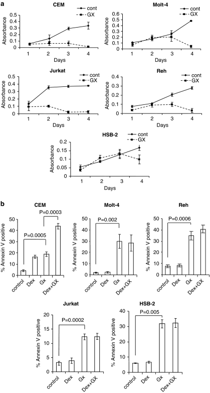
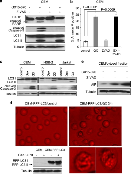
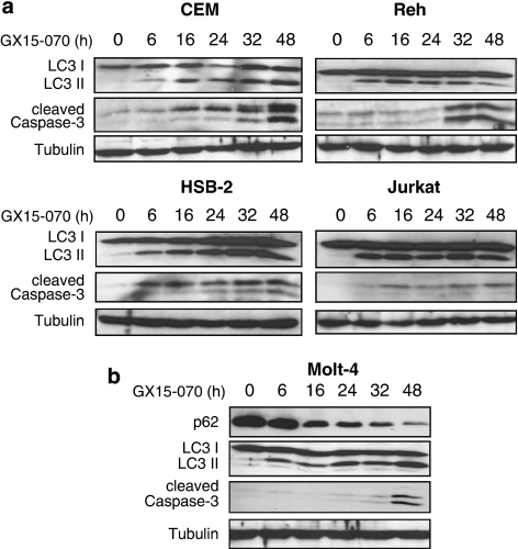
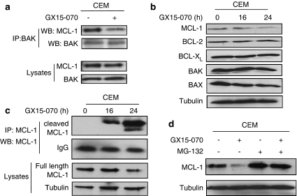
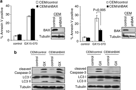
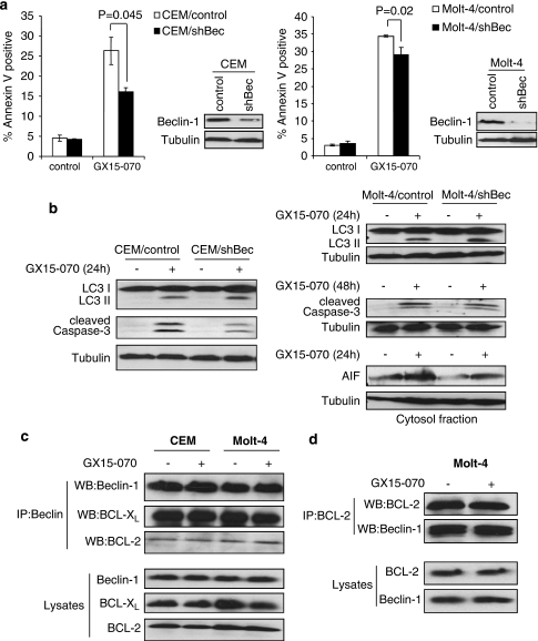
Glucocorticoids (GCs) are common components of many chemotherapeutic regimens for lymphoid malignancies including acute lymphoblastic leukemia (ALL). The BCL-2 family has an essential role in regulating GC-induced cell death. Here we show that downregulation of antiapoptotic BCL-2 family proteins, especially MCL-1, enhances GC-induced cell death. Thus we target MCL-1 by using GX15-070 (obatoclax) in ALL cells. Treatment with GX15-070 in both dexamethasone (Dex)-sensitive and -resistant ALL cells shows effective growth inhibition and cell death. GX15-070 induces caspase-3 cleavage and increases the Annexin V-positive population, which is indicative of apoptosis. Before the onset of apoptosis, GX15-070 induces LC3 conversion as well as p62 degradation, both of which are autophagic cell death markers. A pro-apoptotic molecule BAK is released from the BAK/MCL-1 complex following GX15-070 treatment. Consistently, downregulation of BAK reduces caspase-3 cleavage and cell death, but does not alter LC3 conversion. In contrast, downregulation of ATG5, an autophagy regulator, decreases LC3 conversion and cell death, but does not alter caspase-3 cleavage, suggesting that apoptosis and autophagy induced by GX15-070 are independently regulated. Downregulation of Beclin-1, which is capable of crosstalk between apoptosis and autophagy, affects GX15-070-induced cell death through apoptosis but not autophagy. Taken together, GX15-070 treatment in ALL could be an alternative regimen to overcome glucocorticoid resistance by inducing BAK-dependent apoptosis and ATG5-dependent autophagy.


Figures

Figure 1
Figure 1
Downregulation of MCL-1 enhances dexamethasone-induced lethality. Left panel: CEM cells were infected with lentiviruses expressing shRNAs for non-targeting control, BIM, MCL-1, or BCL-2. Puromycin-resistant cells were pooled after each infection. Equal amounts of total cell extracts were subjected to western blotting with the indicated antibodies. Right panel: Cells were treated with 100 nM Dex for 48 h. The percentage of apoptotic cells was determined by Annexin V-propidium iodide (PI) staining followed by FACS analysis. Values represent the mean±S.D. of three independent experiments
Figure 2
Figure 2
GX15-070 has lethality effect in both Dex-sensitive and -resistant ALL cells. (a) ALL cell lines including CEM, Molt-4, Jurkat, Reh, or HSB-2 were treated with 100 nM GX15-070 (GX) for indicated times and growth rate was determined by WST-1 colorimetric assay. Error bars represent the standard error of three independent experiments. (b) Cells were treated with 100 nM Dex and/or 100 nM GX15-070 for 48 h. The percentage of cell death was determined by Annexin V-PI staining followed by FACS analysis. Values represent the mean±S.D. of three independent experiments
Figure 3
Figure 3
Apoptosis and autophagy are induced by GX15-070 treatment. (a) CEM cells were treated with 100 nM GX15-070±50 μM Z-VAD for 32 h. Z-VAD was added 30 min before GX15-070 treatment. Cleavage of PARP or caspase-3 and LC3 conversion were detected by western blot analyses. (b) CEM cells were treated with 100 nM GX15-070 in the presence or absence of 50 μM Z-VAD for 48 h. The percentage of cell death was determined by Annexin V-PI staining followed by FACS analysis. Values represent the mean±S.D. of three independent experiments. (c) Dex-sensitive CEM or -resistant HSB-2 and Jurkat cells were treated with 100 nM Dex or 100 nM GX15-070 for 24 h, and LC3 conversion and caspase-3 cleavage were detected by western blot analyses. (d) CEM cells were stably transfected with RFP-LC3, and G418-resistant clone cells were then treated with 100 nM GX15-070 for 24 h. Punctated RFP-LC3 was visualized under a fluorescent microscope. Western blot analysis confirmed the conversion of RFP-LC3. These pictures are representative of three independent experiments. Magnification is × 40. Bottom pictures are enlarged single-cell images. (e) CEM cells were treated with 100 nM GX15-070±50 μM Z-VAD for 24 h and release of cytochrome c or AIF in cytosol fraction was determined by western blot analyses
Figure 4
Figure 4
Autophagy is initiated before apoptosis by GX15-070 treatment. (a) CEM, Reh, HSB-2, or Jurkat cells were treated with 100 nM GX15-070 for the indicated times. LC3 conversion and caspase-3 cleavage were determined by western blot analyses. (b) Molt-4 cells were treated with 100 nM GX15-070 for the indicated times. LC3 conversion and degradation of p62 as well as caspase-3 cleavage were demonstrated by western blot analyses
Figure 5
Figure 5
GX15-070 induces dissociation of MCL-1/BAK complex. (a) CEM cells were treated with 100 nM GX15-070 for 5 h. Total cell extracts were subjected to immunoprecipitation with anti-BAK. Western blotting was performed on precipitated samples and on lysates collected before immunoprecipitation with MCL-1 or BAK antibodies. (b) CEM cells were treated with 100 nM GX15-070 for the indicated times and the levels of MCL-1, BCL-2, BCL-XL, BAX, and BAK were examined by western blot analysis. (c) CEM cells were treated with 100 nM GX15-070 for the indicated times. Total cell extracts were subjected to immunoprecipitation with anti-MCL-1. Western blotting was performed on precipitated samples and on lysates collected before immunoprecipitation with anti-MCL-1. (d) CEM cells were treated with 100 nM GX15-070 for 24 h in the presence or absence of 500 nM MG-132. The level of Mcl-1 was determined by western blot analysis
Figure 6
Figure 6
GX15-070-induced apoptosis is BAK-dependent. (a) CEM cells were infected with lentiviruses expressing shRNAs for non-targeting control, BAX, or BAK. Puromycin-resistant cells were pooled after each infection. Downregulation of BAX or BAK expression was monitored by western blotting (in sets). Cells were treated with 100 nM GX15-070 for 48 h and the percentage of cell death was determined by Annexin V-PI staining followed by FACS analysis. Values represent the mean±S.D. of three independent experiments. (b) Cells harboring control, BAX, or BAK shRNA were treated with 100 nM GX15-070 for 32 h. Caspase-3 cleavage and LC3 conversion was determined by western blot analysis
Figure 7
Figure 7
Downregulation of ATG5 decreases autophagy-mediated cell death induced by GX15-070 treatment. (a) Molt-4 cells were infected with lentiviruses expressing shRNAs for either non-targeting control or ATG5. Puromycin-resistant cells were pooled after each infection. Downregulation of ATG5 mRNA was monitored by q-PCR (bottom panel). Cells were treated with 100 nM GX15-070 for 72 h and the percentage of cell death was determined by Annexin V-PI staining followed by FACS analysis. Values represent the mean±S.D. of three independent experiments. (b) Cells were treated with 100 nM GX15-070 for the indicated times, and LC3 conversion and caspase-3 cleavage was assessed by western blot analyses
Figure 8
Figure 8
Beclin-1 contributes to GX15-070-induced apoptosis but not to autophagy. (a) CEM or Molt-4 cells were infected with lentiviruses expressing shRNAs for either non-targeting control or Beclin-1 (Bec). Puromycin-resistant cells were pooled after each infection. Downregulation of Beclin-1 expression was monitored by western blotting (in sets). Cells were treated with 100 nM GX15-070 for 48 h and the percentage of cell death was determined by Annexin V-PI staining followed by FACS analysis. Values represent the mean±S.D. of three independent experiments. (b) Cells were treated with 100 nM GX15-070 for the indicated times, and LC3 conversion, caspase-3 cleavage, and AIF release were assessed by western blot analyses. (c) CEM or Molt-4 cells were treated with 100 nM GX15-070 for 24 h. Total cell extracts were subjected to immunoprecipitation with anti-Beclin-1. Western blotting was performed on precipitated samples and on lysates collected before immunoprecipitation with Beclin-1, BCL-2, or BCL-XL antibodies. (d) Reciprocal immunoprecipitation was performed with anti-BCL-2. Western blotting was conducted on precipitated samples and on lysates collected before immunoprecipitation with BCL-2 or Beclin-1 antibodies

References

    1. Tissing WJ, Meijerink JP, den Boer ML, Pieters R. Molecular determinants of glucocorticoid sensitivity and resistance in acute lymphoblastic leukemia. Leukemia. 2003;17:17–25. - PubMed
    1. Schmidt S, Rainer J, Ploner C, Presul E, Riml S, Kofler R. Glucocorticoid-induced apoptosis and glucocorticoid resistance: molecular mechanisms and clinical relevance. Cell Death Differ. 2004;11 (Suppl 1:S45–S55. - PubMed
    1. Bachmann PS, Gorman R, Papa RA, Bardell JE, Ford J, Kees UR, et al. Divergent mechanisms of glucocorticoid resistance in experimental models of pediatric acute lymphoblastic leukemia. Cancer Res. 2007;67:4482–4490. - PubMed
    1. Tissing WJ, Meijerink JP, Brinkhof B, Broekhuis MJ, Menezes RX, den Boer ML, et al. Glucocorticoid-induced glucocorticoid-receptor expression and promoter usage is not linked to glucocorticoid resistance in childhood ALL. Blood. 2006;108:1045–1049. - PubMed
    1. Distelhorst CW. Recent insights into the mechanism of glucocorticosteroid-induced apoptosis. Cell Death Differ. 2002;9:6–19. - PubMed

Publication types

MeSH terms