. 2011 Feb 18;6(2):e16967.
doi: 10.1371/journal.pone.0016967.

Localized delivery of interferon-β by Lactobacillus exacerbates experimental colitis

Affiliations

Localized delivery of interferon-β by Lactobacillus exacerbates experimental colitis

Adelle P McFarland et al. PLoS One. .

Abstract

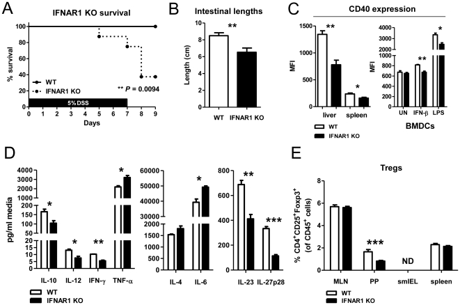
Background: There have been conflicting reports of the role of Type I interferons (IFN) in inflammatory bowel disease (IBD). Clinical trials have shown potent efficacy of systemic interferon-beta (IFN-β) in inducing remission of ulcerative colitis. Likewise, IFNAR1(-/-) mice display an increased sensitivity to dextran sulfate sodium (DSS)-induced colitis, suggesting Type I IFN play a protective role during inflammation of the gut. Curiously, however, there have also been reports detailing the spontaneous development of IBD in patients receiving systemic IFN-β therapy for multiple sclerosis or hepatitis.

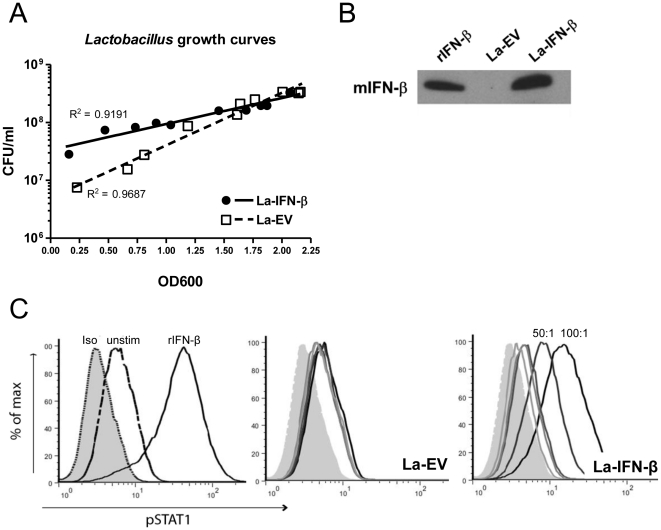
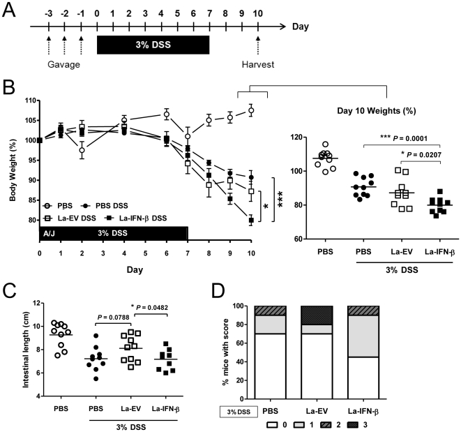
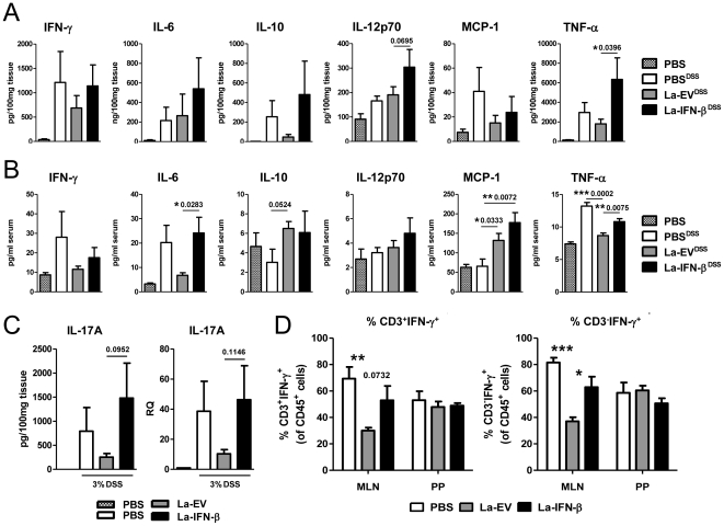
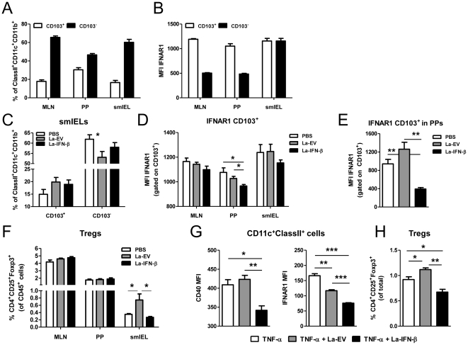
Methodology/principal findings: To investigate the effects of local administration of IFN-β on a murine model of colitis, we developed a transgenic Lactobacillus acidophilus strain that constitutively expresses IFN-β (La-IFN-β). While pretreatment of mice with control Lactobacillus (La-EV) provided slight protective benefits, La-IFN-β increased sensitivity to DSS. Analysis showed colitic mice pretreated with La-IFN-β had increased production of TNF-α, IFN-γ, IL-17A and IL-13 by intestinal tissues and decreased regulatory T cells (Tregs) in their small intestine. Examination of CD103(+) dendritic cells (DCs) in the Peyer's patches revealed that IFNAR1 expression was dramatically reduced by La-IFN-β. Similarly, bone marrow-derived DCs matured with La-IFN-β experienced a 3-fold reduction of IFNAR1 and were impaired in their ability to induce Tregs.

Conclusions/significance: Our IFNAR1 expression data identifies a correlation between the loss/downregulation of IFNAR1 on DCs and exacerbation of colitis. Our data show that Lactobacillus secreting IFN-β has an immunological effect that in our model results in the exacerbation of colitis. This study underscores that the selection of therapeutics delivered by a bacterial vehicle must take into consideration the simultaneous effects of the vehicle itself.


Conflict of interest statement

Competing Interests: The authors have declared that no competing interests exist.

Figures

Figure 1
Figure 1. IFNAR1−/− mice are more susceptible to DSS-induced colitis and have altered DC and Treg profiles.
(A, B) IFNAR1−/− mice experienced increased weight loss and greater degree of intestinal shortening when administered 5% DSS in their drinking water for 7 d compared to wild-type controls (n = 10). (C) DCs isolated from liver and spleen of IFNAR1−/− mice and BMDCs matured with IFN-β (1000 U/ml) or LPS (100 ng/ml) for 24 h were analyzed for cell surface expression of CD40. DCs were gated as CD45+ClassII+CD11c+. (D) Levels of cytokines, measured by CBA or ELISA (IL-23 and IL-27p28), in BMDC supernatants stimulated with LPS (100 ng/ml) for 24 h from IFNAR1−/− or wild-type mice (n = 5). (E) Percent Tregs, gated as CD45+CD4+CD25+Foxp3+, in MLNs, PPs, smIELs and spleens of IFNAR1−/− and wild-type mice (ND = not detected, n = 10). The data is represented as the mean ± SEM. * p<0.05, ** p<0.01, *** p<0.001.
Figure 2
Figure 2. La-IFN-β expresses biologically functional murine IFN-β.
(A) Growth curves for La-IFN-β and La-EV strains correlating CFU/ml of bacterial culture with OD600 values. (B) Western blot showing detection of murine IFN-β in the supernatant from an overnight culture of transgenic La-IFN-β. Prior to Western blotting, the protein was precipitated using TCA. Positive control is 100 U of recombinant murine IFN-β. (C) Phosphorylation of STAT1 in a C57BL/6 macrophage cell line after 60 min stimulation with rIFN-β (1000 U) as a positive control or co-culture with 5∶1, 10∶1, 50∶1 or 100∶1 ratios of La-EV or La-IFN-β.
Figure 3
Figure 3. Pretreatment of mice with La-IFN-β exacerbates DSS-induced colitis.
(A) On days -3, -2 and -1 mice were gastric gavaged with 109 CFU of the bacterial treatments and days 0-7 3% DSS was administered in the drinking water. (B) Percent body weight loss of mice pretreated with La-IFN-β, La-EV or PBS for three days. The 100% body weights were taken as the weights of mice just prior to DSS administration. Day 10 percent body weights are plotted to show individual mice (n = 10). (C) Intestinal lengths (cm) of mice on day 10 following DSS administration. Colons were harvested and measured from the proximal end just below the cecum and to the anus (n = 10). (D) Following the harvest of the colons, the tissue was macroscopically examined for severity and distribution of intestinal hemorrhaging and scored on a scale from 0–3 (0 = no visible hemorrhaging, 1 = diffuse hemorrhaging, 2 = widespread hemorrhaging, 3 = severe hemorrhaging throughout colon). Results shown are representative of three independent experiments.
Figure 4
Figure 4. Elevated levels of inflammatory cytokines in mice pretreated with La-IFN-β over La-EV.
(A) 2 cm of the distal colon was removed and weighed from colitic mice pretreated with PBS, La-EV or La-IFN-β (and a no DSS control group) and cultured overnight (n = 10). IFN-γ, IL-6, IL-10, IL-12p70, MCP-1 and TNF-α cytokine levels were assayed by CBA and standardized to concentration per 100 mg tissue. (B) Serum was assayed for the presence of inflammatory cytokines by CBA on day 7 of DSS administration (n = 5). (C) Measurement of IL-17A in colonic tissue culture (as outlined in Fig.4A) by CBA and the relative quantification of IL-17A mRNA in the distal colon was measured by RT-PCR. For RT-PCR, the colitic groups were compared relative to a no DSS control (RQ = 1) and HRPT was used as an endogenous control. (D) Cells isolated from the MLNs and PPs of colitic mice were stimulated with PMA (10 ng/ml) and ionomycin (1 µM/ml) for 5 h with the addition of Brefeldin A after 2 h. The cells were then stained for CD3 and fixed for intracellular detection of IFN-γ. Data is presented as %CD3+IFN-γ+ or %CD3IFN-γ+ cells in the CD45+ population. The data is represented as the mean ± SEM. * p<0.05, ** p<0.01, *** p<0.001.
Figure 5
Figure 5. IFNAR1 is decreased on CD103+ DCs in the PPs of mice following treatment with La-IFN-β.
(A) Percent distribution of CD103+ versus CD103 DCs in the MLNs, PPs and smIELs of wild-type mice (n = 5) gated on CD45+ClassII+CD11c+CD11b+ cells. (B) IFNAR1 expression (mean fluorescence intensity, MFI) on CD103+ and CD103 DCs in the MLNs, PPs and smIELs from wild-type mice. (C) CD103+ and CD103 DC percentages in the smIELs of colitic mice pretreated with PBS, La-EV or La-IFN-β (n = 5). (D) IFNAR1 expression (MFI) on CD103+ DCs in the MLNs, PPs and smIELs of colitic mice in the different treatment groups. (E) IFNAR1 expression on CD103+ DCs in the PPs of healthy mice following three consecutive days of gastric gavage with PBS, La-EV or La-IFN-β. (F) Cells were isolated from colitic mice pretreated with PBS, La-EV or La-IFN-β (n = 5) from the MLNs, PPs and smIELs and stained for the presence of Tregs, which were gated as CD45+CD4+CD25+Foxp3+. The graph shows the % Tregs present in the CD45+ population. (G, H) BMDCs from wild-type mice were matured with TNF-α (50 ng/ml), TNF-α + La-EV or TNF-α+ La-IFN-β (100∶1 ratio, bacteria:BMDCs) for 2 d. The mature BMDCs were then co-cultured with CD3/CD28 (1 µg/ml) activated splenocytes for 7 d (1∶2 ratio, BMDCs:splenocytes). Following co-culture, DCs (gated as ClassII+CD11c+) were stained for CD40 and IFNAR1 expression (G) and the percent Tregs (CD4+CD25+Foxp3+) was determined (H). The data is represented as the mean ± SEM. * p<0.05, ** p<0.01, *** p<0.001.

References

    1. Platanias LC. Mechanisms of type-I- and type-II-interferon-mediated signalling. Nat Rev Immunol. 2005;5:375–386. - PubMed
    1. Benveniste EN, Qin H. Type I interferons as anti-inflammatory mediators. Sci STKE 2007. 2007;416:pe70. - PubMed
    1. Billiau A. Anti-inflammatory properties of Type I interferons. Antiviral Res. 2006;71:108–116. - PMC - PubMed
    1. van HJ, Reedquist K, Sattonet-Roche P, Smeets TJ, Plater-Zyberk C, et al. Treatment with recombinant interferon-beta reduces inflammation and slows cartilage destruction in the collagen-induced arthritis model of rheumatoid arthritis. Arthritis Res Ther. 2004;6:R239–R249. - PMC - PubMed
    1. Triantaphyllopoulos KA, Williams RO, Tailor H, Chernajovsky Y. Amelioration of collagen-induced arthritis and suppression of interferon-gamma, interleukin-12, and tumor necrosis factor alpha production by interferon-beta gene therapy. Arthritis Rheum. 1999;42:90–99. - PubMed

Publication types

MeSH terms