Comparative Study
. 2011 Mar 2;31(9):3225-33.
doi: 10.1523/JNEUROSCI.2568-10.2011.

Axonally derived neuregulin-1 is required for remyelination and regeneration after nerve injury in adulthood

Affiliations
Comparative Study

Axonally derived neuregulin-1 is required for remyelination and regeneration after nerve injury in adulthood

Florence R Fricker et al. J Neurosci. .

Abstract

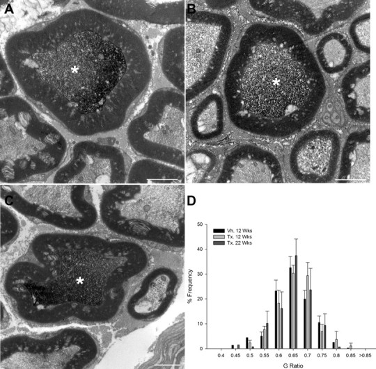
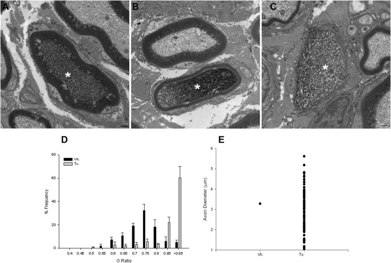
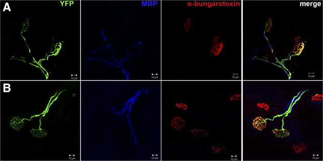
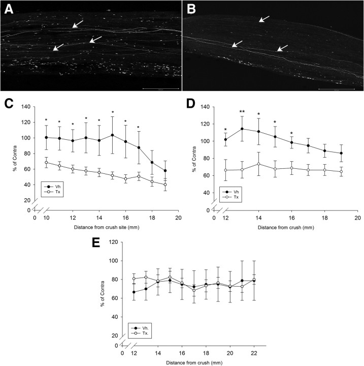
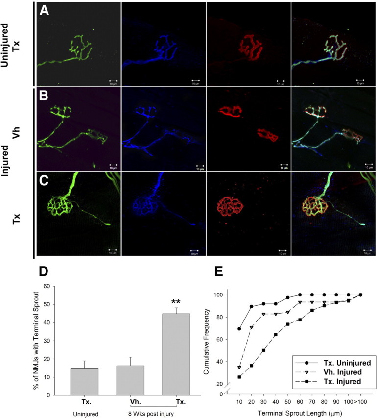
Neuregulin-1 (NRG1) plays a crucial role in axoglial signaling during the development of the peripheral nervous system, but its importance in adulthood after peripheral nerve injury remains unclear. We used single-neuron labeling with inducible Cre-mediated knock-out animals, which enabled visualization of a subset of adult myelinated sensory and motoneurons neurons in which Nrg1 was inducibly mutated by tamoxifen treatment. In uninjured mice, NRG1-deficient axons and the associated myelin sheath were normal, and the neuromuscular junction demonstrated normal apposition of presynaptic and postsynaptic components. After sciatic nerve crush, NRG1 ablation resulted in severe defects in remyelination: axons were either hypomyelinated or had no myelin sheath. NRG1-deficient axons were also found to regenerate at a slower rate. After nerve injury, the neuromuscular junction was reinnervated, but excess terminal sprouting was observed. Juxtacrine Neuregulin-1 signaling is therefore dispensable for maintenance of the myelin sheath in adult animals but has a key role in reparative processes after nerve injury.


Figures

Figure 1.
Figure 1.
A schematic representation of the sciatic nerve crush model used to assess axon remyelination and regeneration. Tamoxifen (Tamox.) or vehicle (Veh.) was administered to animals of 10 weeks of age for 5 d. At 4 weeks later, the sciatic nerve was crushed at the midthigh level. At 10 d, 14 d, or 8 weeks after sciatic nerve crush, this nerve was harvested at a point 2 mm distal to the crush site and processed for immunoelectron microscopy. A whole-mount preparation of the tibial nerve was developed to visualize regenerating YFP-positive axons. The gastrocnemius muscle (which is innervated by the tibial nerve) was used for studies of neuromuscular junction reinnervation.
Figure 2.
Figure 2.
NRG1 ablation is specific to YFP-expressing motoneurons and large DRG cells after tamoxifen treatment. A, In situ hybridization images using a probe directed against the βEGF domain of NRG1 in L4 DRG. B, In situ hybridization images of the ventral horn of the spinal cord at the level of the lumbar enlargement. Images are of tamoxifen-dosed and vehicle-dosed SLICK-A Cre; NRG-1fl/fl animals (conditional NRG1 mutant). A population of motoneurons within the ventral horn of the spinal cord and a small population of DRG cells express YFP (arrows). In tamoxifen-dosed animals, NRG1 expression is absent in these cells (whereas in YFP-negative cells, expression of NRG1 is maintained). Vh, Vehicle; Tx, tamoxifen. Scale bars, 50 μm.
Figure 3.
Figure 3.
Juxtacrine NRG1 signaling is dispensable for maintenance of the myelin sheath in uninjured adult animals. In SLICK-A animals, YFP-expressing axons can be identified by preembedding immunoelectron microscopy by the presence of a dark reaction product (asterisks). A–D, There is no change in the morphology of YFP-positive axons within the sciatic nerve at 12 (B) or 22 (C) weeks after administration of tamoxifen to SLICK-A Cre; NRG-1fl/fl mice compared with control animals (12 weeks after vehicle treatment; A). D, G-ratio frequency distribution in nerves of conditional NRG1 mutant and control mice; n = 3–5 per group. Vh., Vehicle; Tx., tamoxifen. Scale bars, 2 μm.
Figure 4.
Figure 4.
Axon-derived NRG1 is required for remyelination after nerve injury in the adult. A, At 8 weeks after sciatic nerve crush, control (vehicle-treated SLICK-A Cre; NRG-1fl/fl) animals demonstrated effective remyelination of YFP-expressing axons. BD, In contrast, YFP-positive axons from conditional NRG1 mutant (tamoxifen-treated SLICK-A Cre; NRG-1fl/fl) mice either had a significantly thinner myelin sheath (B) or completely failed to elaborate a myelin sheath (C; quantified in D, G-ratio plot; p < 0.001, Kolmogorov–Smirnov test). Note in B a neighboring axon that is YFP negative is normally myelinated. E, Graphic plot displaying numbers of unmyelinated YFP-positive axons (n = 4 animals per group). Vh., Vehicle; Tx., tamoxifen. Scale bar, 500 nm.
Figure 5.
Figure 5.
NRG1-deficient axons could be visualized in the gastrocnemius muscle with no myelin sheath 8 weeks after sciatic nerve crush. A, In SLICK-A Cre; NRG-1fl/fl animals, regenerated motor axons (YFP, green) at 8 weeks after sciatic nerve crush that have reinnervated an α-bungarotoxin (red)-labeled NMJ can be visualized. MBP staining (blue) colocalizes with YFP staining. B, NMJ from tamoxifen-treated SLICK-A Cre; NRG-1fl/fl animal (i.e., conditional NRG1 mutant); MBP staining is frequently absent over long lengths from YFP-labeled axons, despite neighboring non-YFP axons staining for MBP. Scale bar, 10 μm.
Figure 6.
Figure 6.
NRG1-deficient axons regenerate at a slower rate. A, B, YFP-positive axons (see arrows) within the full thickness of the tibial nerve can be visualized using confocal microscopy 10 d after sciatic nerve crush in vehicle-treated (A) and tamoxifen-treated (B) SLICK-A Cre; NRG-1fl/fl animals. Note the smaller number of regenerating axons in the conditional NRG1 mutant (i.e., tamoxifen-treated SLICK-A Cre; NRG-1fl/fl). This is quantified in C–E, expressed as a percentage of axons relative to the contralateral uncrushed side versus distance from crush site. There is a significant difference between the tamoxifen- and vehicle-treated group at both 10 (C) and 14 (D) days after crush (n = 7 per group; *p < 0.05, **p < 0.005, two-way ANOVA post hoc Tukey's test). E, At 2 months after crush, axons had effectively regenerated through the tibial nerve in both groups, with no significant difference between them (n = 5 per group). Vh., Vehicle; Tx., tamoxifen. Scale bars, 500 μm.
Figure 7.
Figure 7.
NRG1 ablation results in excess terminal sprouting at the neuromuscular junction after peripheral nerve injury. A, The neuromuscular junction 12 weeks after tamoxifen administration to SLICK-A Cre; NRG-1fl/fl animals (conditional NRG1 mutant) demonstrated normal apposition and morphology of presynaptic and postsynaptic elements (YFP, green; S100, blue; α-bungarotoxin, red, respectively). B, C, At 8 weeks after sciatic nerve crush; C, NMJs of conditional NRG1 mutant animals were reinnervated but had a much higher degree of terminal sprouting compared with control (B). These axonal sprouts clearly followed the processes of terminal Schwann cells immunolabeled by S-100 (blue). D, There was a significant increase in the proportion of NMJs with sprouts (**p < 0.005, one-way ANOVA post hoc Tukey's test). E, When terminal sprout length is plotted as cumulative sum distributions, sciatic crush results in a small but significant increase in sprout length in vehicle-treated animals (p < 0.005, Kolmogorov–Smirnov test), but sprout length is significantly greater in tamoxifen-treated animals (p < 0001, Kolmogorov–Smirnov test). n = 3 animals per group. Vh., Vehicle; Tx., tamoxifen. Scale bars, 10 μm.

Similar articles

Cited by

References

    1. Atanasoski S, Scherer SS, Sirkowski E, Leone D, Garratt AN, Birchmeier C, Suter U. ErbB2 signaling in Schwann cells is mostly dispensable for maintenance of myelinated peripheral nerves and proliferation of adult Schwann cells after injury. J Neurosci. 2006;26:2124–2131. - PMC - PubMed
    1. Baloh RH, Strickland A, Ryu E, Le N, Fahrner T, Yang M, Nagarajan R, Milbrandt J. Congenital hypomyelinating neuropathy with lethal conduction failure in mice carrying the Egr2 I268N mutation. J Neurosci. 2009;29:2312–2321. - PMC - PubMed
    1. Bao J, Wolpowitz D, Role LW, Talmage DA. Back signaling by the Nrg-1 intracellular domain. J Cell Biol. 2003;161:1133–1141. - PMC - PubMed
    1. Bermingham-McDonogh O, Xu YT, Marchionni MA, Scherer SS. Neuregulin expression in PNS neurons: isoforms and regulation by target interactions. Mol Cell Neurosci. 1997;10:184–195. - PubMed
    1. Brinkmann BG, Agarwal A, Sereda MW, Garratt AN, Müller T, Wende H, Stassart RM, Nawaz S, Humml C, Velanac V, Radyushkin K, Goebbels S, Fischer TM, Franklin RJ, Lai C, Ehrenreich H, Birchmeier C, Schwab MH, Nave KA. Neuregulin-1/ErbB signaling serves distinct functions in myelination of the peripheral and central nervous system. Neuron. 2008;59:581–595. - PMC - PubMed

Publication types

MeSH terms