Comparative Study
. 2011 Mar 2;31(9):3290-4.
doi: 10.1523/JNEUROSCI.6256-10.2011.

Improving visual sensitivity with subthreshold transcranial magnetic stimulation

Affiliations
Comparative Study

Improving visual sensitivity with subthreshold transcranial magnetic stimulation

Arman Abrahamyan et al. J Neurosci. .

Abstract

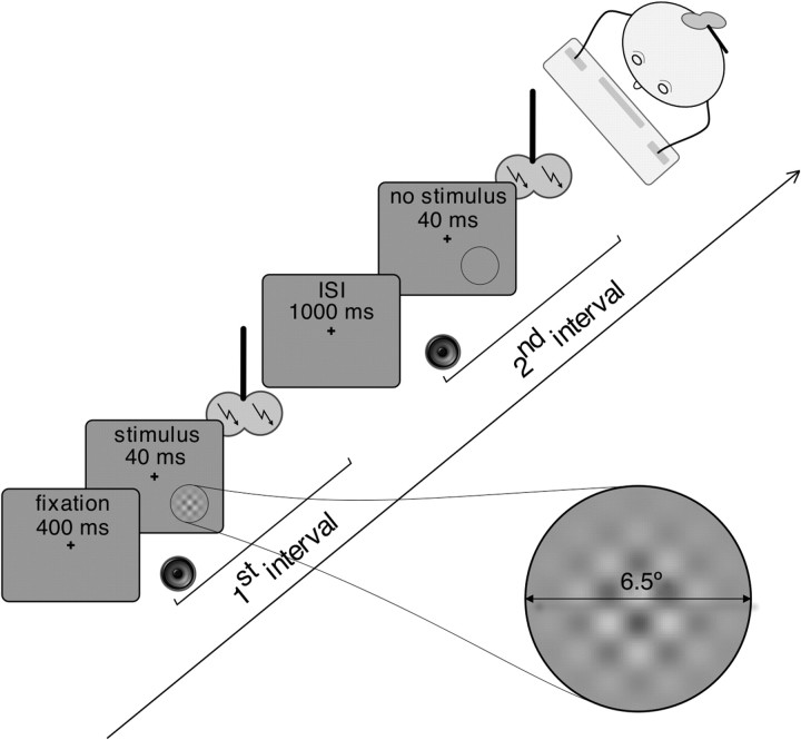
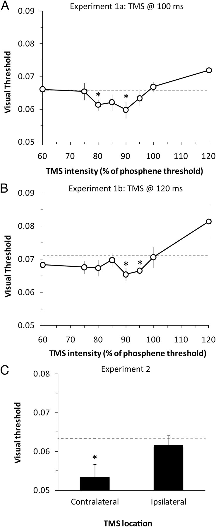
We probed for improvement of visual sensitivity in human participants using transcranial magnetic stimulation (TMS). Stimulation of visual cortex can induce an illusory visual percept known as a phosphene. It is known that TMS, delivered at intensities above the threshold to induce phosphenes, impairs the detection of visual stimuli. We investigated how the detection of a simple visual stimulus is affected by TMS applied to visual cortex at or below the phosphene threshold. Participants performed the detection task while the contrast of the visual stimulus was varied from trial to trial according to an adaptive staircase procedure. Detection of the stimulus was enhanced when a single pulse of TMS was delivered to the contralateral visual cortex 100 or 120 ms after stimulus onset at intensities just below the phosphene threshold. No improvement in visual sensitivity was observed when TMS was applied to the visual cortex in the opposite hemisphere (ipsilateral to the visual stimulus). We conclude that TMS-induced neuronal activity can sum with stimulus-evoked activity to augment visual perception.


Figures

Figure 1.
Figure 1.
Experiment design. In three experiments, we tested participants on a two-interval, two-alternative forced-choice task. A visual plaid stimulus was presented for 40 ms in the middle of one of two sequential 1 s intervals (first or second interval, with equal probability). The start of each interval was signaled by a brief auditory cue. Participants had to fixate the cross in the center of the monitor and report which interval contained the visual stimulus by pressing one of two keys. During both intervals, a single pulse of TMS was delivered either to the occipital lobe (experimental conditions) or to the Cz site located on the top of the head at the intersection of the midline and interaural line (control conditions).
Figure 2.
Figure 2.
A–C, Detection thresholds, relative to the Cz control condition (dashed black line), in experiments 1a (A), 1b (B), and 2 (C). Values below the dashed line indicate an improvement in visual sensitivity. In experiments 1a and 1b, a single pulse of TMS was delivered to visual cortex 100 or 120 ms after onset of the visual stimulus. The intensity of TMS varied from 60 to 120% of the phosphene threshold. Experiment 2 compared the effect of TMS applied to visual cortex contralateral or ipsilateral to the stimulus, 120 ms after stimulus onset at an intensity just below phosphene threshold. Error bars show within-subject SEM (for the difference with Cz), and asterisks identify thresholds that were significantly different from Cz (p < 0.05).
Figure 3.
Figure 3.
A illustrates a nonlinear relationship between input strength (stimulation intensity) and response strength in a sensory system. The horizontal gridlines mark the smallest resolvable units of response strength (that are perceptually discriminable); the spacing between the vertical gridlines shows the smallest detectable change in input strength (the JND). Td marks the minimum detectable input strength (the absolute detection threshold). Psychophysical evidence that the relationship between sensory input and response strength follows a sigmoid function, as shown here, comes from demonstrations that the JND between two stimuli decreases as their base strength increases, when the base strength is small (close to Td), but beyond this point the JND increases as the base strength increases, as described by Weber's Law (Solomon, 2009). The biphasic nature of this change in JND as a function of base stimulus strength produces a dipper function, as shown in B.

References

    1. Allen EA, Pasley BN, Duong T, Freeman RD. Transcranial magnetic stimulation elicits coupled neural and hemodynamic consequences. Science. 2007;317:1918–1921. - PubMed
    1. Amassian VE, Cracco RQ, Maccabee PJ, Cracco JB, Rudell A, Eberle L. Suppression of visual perception by magnetic coil stimulation of human occipital cortex. Electroencephalogr Clin Neurophysiol. 1989;74:458–462. - PubMed
    1. Arabzadeh E, Clifford CW, Harris JA. Vision merges with touch in a purely tactile discrimination. Psychol Sci. 2008;19:635–641. - PubMed
    1. Barker AT, Jalinous R, Freeston IL. Non-invasive magnetic stimulation of human motor cortex. Lancet. 1985a;1:1106–1107. - PubMed
    1. Barker AT, Freeston IL, Jalinous R, Merton PA, Morton HB. Magnetic stimulation of the human brain. J Physiol (Lond) 1985b;369(Suppl):3P.

Publication types

LinkOut - more resources