Comparative Study
. 2011 Mar 2;31(9):3459-69.
doi: 10.1523/JNEUROSCI.3932-10.2011.

Complement protein C1q-mediated neuroprotection is correlated with regulation of neuronal gene and microRNA expression

Affiliations
Comparative Study

Complement protein C1q-mediated neuroprotection is correlated with regulation of neuronal gene and microRNA expression

Marie E Benoit et al. J Neurosci. .

Abstract

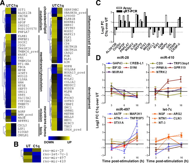
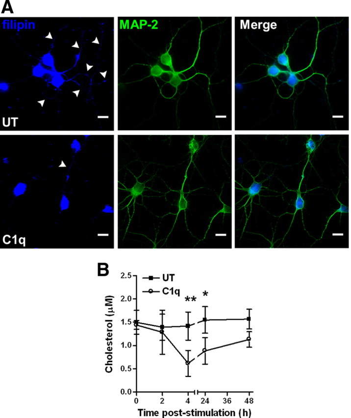
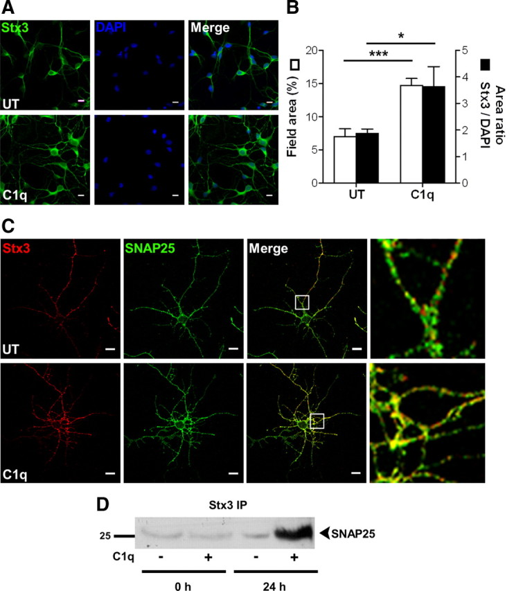
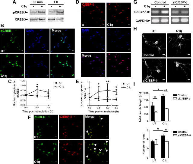
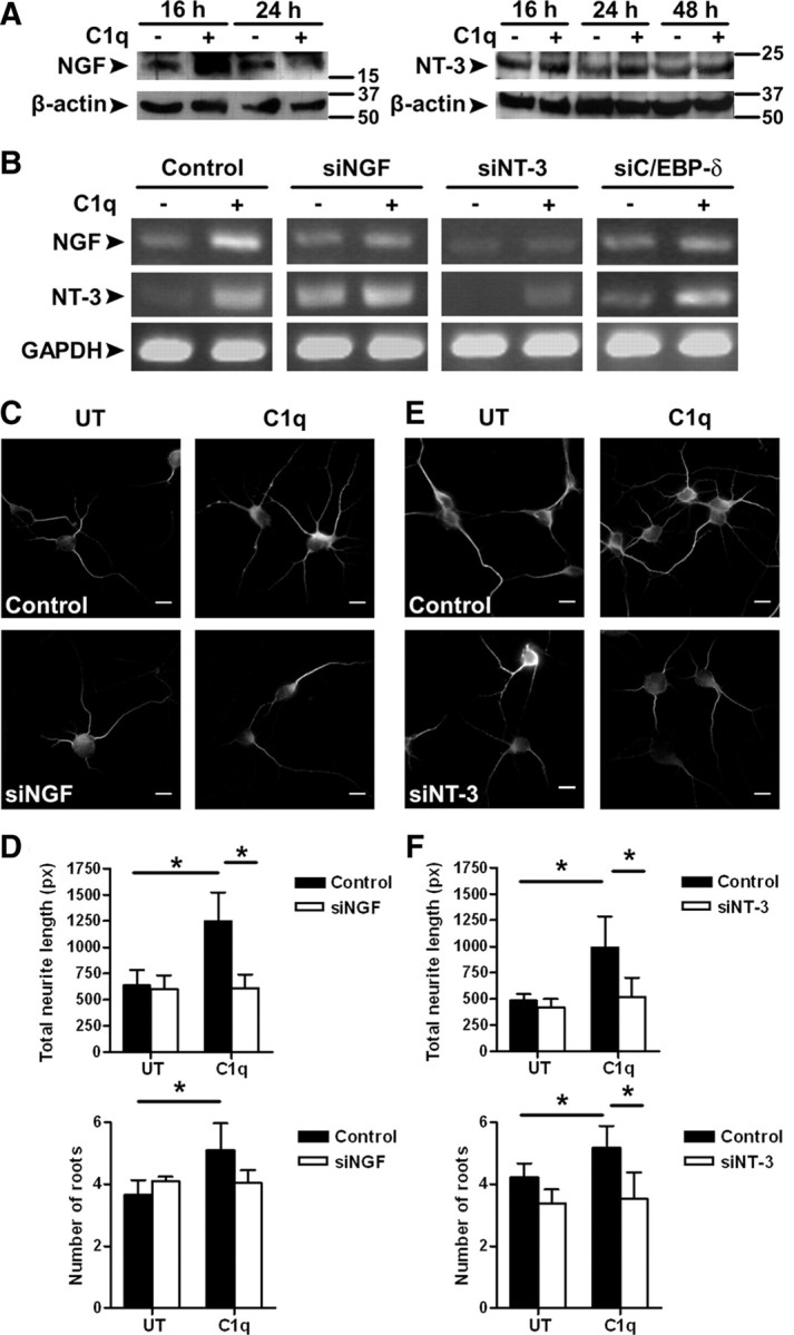
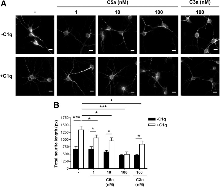
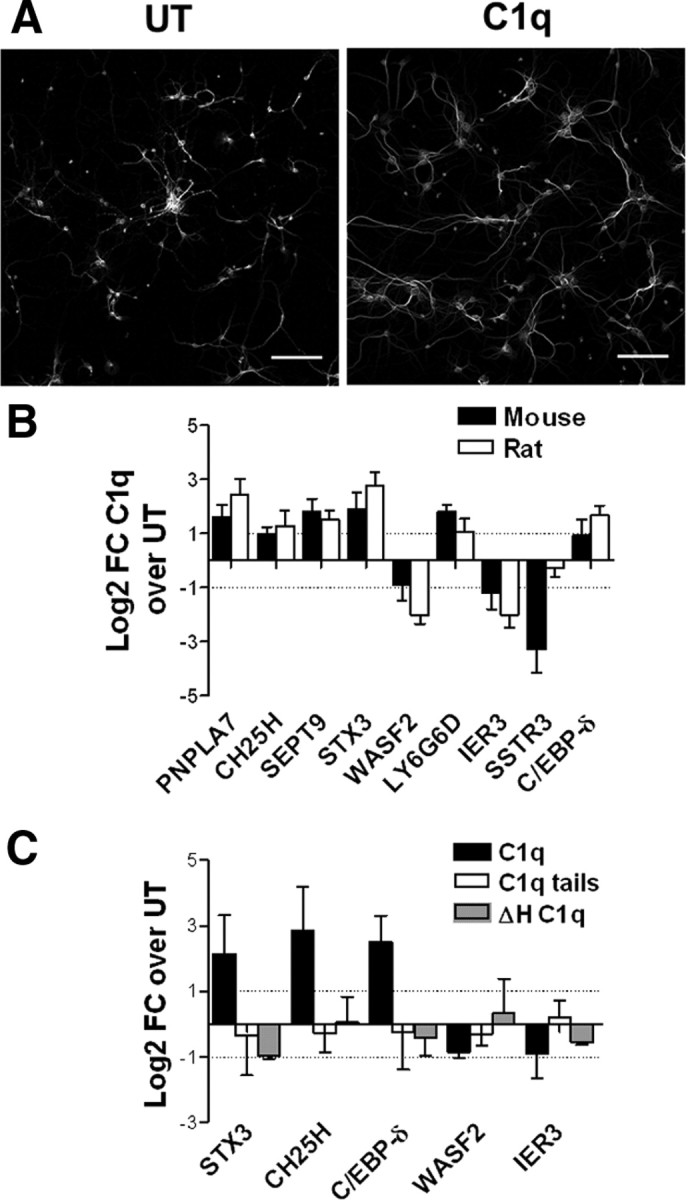
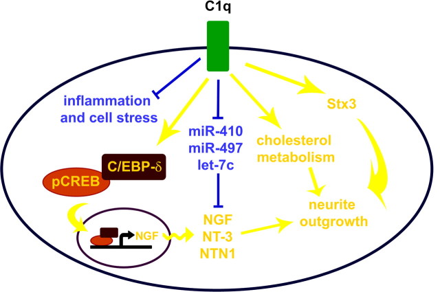
Activation of the complement cascade, a powerful effector mechanism of the innate immune system, is associated with neuroinflammation but also with elimination of inappropriate synapses during development. Synthesis of C1q, a recognition component of the complement system, occurs in brain during ischemia/reperfusion and Alzheimer's disease, suggesting that C1q may be a response to injury. In vitro, C1q, in the absence of other complement proteins, improves neuronal viability and neurite outgrowth and prevents β-amyloid-induced neuronal death, suggesting that C1q may have a direct neuroprotective role. Here, investigating the molecular basis for this neuroprotection in vitro, addition of C1q to rat primary cortical neurons significantly upregulated expression of genes associated with cholesterol metabolism, such as cholesterol-25-hydroxylase and insulin induced gene 2, and transiently decreased cholesterol levels in neurons, known to facilitate neurite outgrowth. In addition, the expression of syntaxin-3 and its functional association with synaptosomal-associated protein 25 was increased. C1q also increased the nuclear translocation of cAMP response element-binding protein and CCAAT/enhancer-binding protein-δ (C/EBP-δ), two transcription factors involved in nerve growth factor (NGF) expression and downregulated specific microRNAs, including let-7c that is predicted to target (and thus inhibit) NGF and neurotrophin-3 (NT-3) mRNA. Accordingly, C1q increased expression of NGF and NT-3, and small interfering RNA inhibition of C/EBP-δ, NGF, or NT-3 expression prevented the C1q-dependent neurite outgrowth. No such neuroprotective effect is seen in the presence of C3a or C5a. Finally, the induced neuronal gene expression required conformationally intact C1q. These results show that C1q can directly promote neuronal survival, thereby demonstrating new interactions between immune proteins and neuronal cells that may facilitate neuroprotection.


Figures

Figure 1.
Figure 1.
Regulation of gene and miRNA expression by C1q in primary cortical neurons. A, B, Functional clustering of GO annotated genes (A) and miRNAs (B) significantly modulated by C1q compared with untreated (UT) neurons. Standardized means shown with a color gradient from blue (downregulated) to yellow (upregulated). C, Comparison of array and qRT-PCR data for 16 significantly modulated genes by C1q. The results are expressed as the means of log2 FC of C1q over untreated. D, Gene expression of miRNA predicted targets studied by qRT-PCR over a 16 h time course study. Results are expressed as the mean ± SD of log2 FC from triplicate samples from two independent experiments. Line color corresponds to the color-coded classifications in supplemental Figure 1 (available at www.jneurosci.org as supplemental material).
Figure 2.
Figure 2.
C1q modifies cholesterol distribution and decreases cholesterol levels in neurons. A, C1q alters cholesterol distribution in neurons. Neurons cultured with or without C1q for 24 h were stained with filipin and anti MAP-2 antibodies and analyzed by fluorescence microscopy. Scale bars, 10 μm. B, Quantification of cholesterol content in untreated (UT) and C1q-treated neurons by enzymatic fluorometric assay over a 48 h time course. Results represent means ± SD (n = 3). Two-way ANOVA followed by Bonferroni's post hoc test, *p < 0.05 and **p < 0.01.
Figure 3.
Figure 3.
C1q increases stx3 expression and its interaction with SNAP25. A, B, C1q increases the protein levels of stx3 in neurons. Neurons cultured with or without C1q for 24 h were stained with anti-stx3. Nuclei are stained with DAPI. A, Representative micrographs of stx3 staining. B, Quantitative image analysis of stx3 expressed as means ± SD (n = 4, 7 fields per condition) of percentage of field area (□) or area ratio stx3/DAPI (■). Two-tailed nonparametric Mann–Whitney U test, *p < 0.05 and ***p < 0.001. C, Colocalization of stx3 and SNAP25 in C1q-treated neurons. Neurons cultured with or without C1q for 24 h were stained with anti-stx3 (Alexa Fluor 555, red) and anti-SNAP25 (Alexa Fluor 488, green) antibodies and analyzed by confocal microscopy. Representative micrographs of two independent experiments are shown with white boxed section expanded on the right. UT, Untreated. Scale bars, 10 μm. D, SNAP25 immunoprecipitates with stx3 after 24 h of stimulation with C1q. One blot representative of three independent experiments is shown. IP, Immunoprecipitation.
Figure 4.
Figure 4.
Signaling through pCREB and C/EBP-δ in C1q-stimulated neurons. A, Expression of pCREB and total CREB in C1q-stimulated neurons assessed by Western blot. One blot representative of three independent experiments is shown. B–E, Nuclear translocation of pCREB and C/EBP-δ in C1q-treated neurons. Neurons treated with C1q for 30 min to 6 h were stained with anti-pCREB (Alexa Fluor 488, green) or anti-C/EBP-δ (Alexa Fluor 555, red) antibodies. Nuclei are stained with DAPI (blue). Representative micrographs of neurons stimulated with C1q for 30 min and stained for pCREB are shown in B. Representative micrographs of neurons stimulated with C1q for 1 h and stained for C/EBP-δ are shown in D. C, E, Quantitative image analysis of nuclear translocation of pCREB (C) and C/EBP-δ (E). Results represent means ± SD (n = 3, 5 fields per condition). Two-way ANOVA, followed by Bonferroni's post hoc test, *p < 0.05 and **p < 0.01. F, Nuclear colocalization of pCREB and C/EBP-δ in C1q-treated neurons after 30 min of stimulation with C1q. Representative micrographs of three independent experiments are shown. Arrows indicate overlapping of pCREB and C/EBP-δ. G–I, C/EBP-δ inhibition prevents C1q-dependent neuroprotection. Knocked down expression of C/EBP-δ after siRNA transfection was assessed by PCR after 6 h of stimulation with C1q (G) and the effect of C/EBP-δ inhibition on C1q-dependent neuroprotection was assessed by MAP-2 immunocytochemistry (H) and image analysis for quantification of total neurite length and number of roots (I) after 24 h of stimulation with C1q. Results represent means ± SD (n = 3, 7 fields per condition). Scale bars, 10 μm. UT, Untreated; px, pixels. Two-tailed nonparametric Mann–Whitney U test, *p < 0.05 and **p < 0.01.
Figure 5.
Figure 5.
Role of neurotrophins in C1q-dependent neuroprotection. A, Expression of NGF and NT-3 in untreated and C1q-treated neurons assessed by Western blot. Representative blots of three independent experiments are shown. B, Expression of NGF, NT-3, and GAPDH after transfection with negative control siRNA or siRNA specific for NGF, NT-3, or C/EBP-δ was assessed by PCR after 6 h of incubation with or without C1q. Gels representative of three independent experiments are shown. C–F, Inhibition of NGF and NT-3 expression prevents C1q-dependent neuroprotection. The effect of NGF and NT-3 inhibition on C1q-dependent neuroprotection was assessed by MAP-2 immunocytochemistry (C, E) and image analysis for quantification of total neurite length and number of roots (D, F) after 24 h of stimulation with C1q. Results represent means ± SD (n = 3, 7 fields per condition). Scale bars, 10 μm. UT, Untreated; px, pixels. Two-tailed nonparametric Mann–Whitney U test, *p < 0.05.
Figure 6.
Figure 6.
C3a and C5a have no direct neuroprotective effect and reduce the neuroprotective effect of C1q. The effect of C3a and C5a on C1q-dependent neuroprotection was assessed by MAP-2 immunocytochemistry (A) and image analysis for quantification of total neurite length (B) after 24 h of stimulation. Results represent means ± SD (n = 3, 5 fields per condition). Scale bars, 10 μm. px, Pixels. Two-tailed nonparametric Mann–Whitney U test, *p < 0.05 and ***p < 0.001.
Figure 7.
Figure 7.
C1q modulates mouse neuronal survival and gene expression. A, C1q promotes neurite outgrowth in primary cortical mouse neurons. Mouse neurons were incubated with or without C1q for 24 h, fixed, and stained with MAP-2 antibodies. Scale bars, 100 μm. B, Mouse and rat primary cortical neurons were incubated with or without C1q for 3 h, and gene expression was assessed by qRT-PCR. C, Mouse primary cortical neurons were incubated with 10 nm C1q, C1q tails, or heat-inactivated C1q (ΔHC1q) for 3 h, and gene expression was assessed by qRT-PCR. Results are expressed as the mean ± SD of log2 FC from triplicate samples from two independent experiments. UT, Untreated.
Figure 8.
Figure 8.
Pathways modulated by C1q in neurons. C1q rapidly upregulates the expression of genes associated with membrane and cytoskeleton function, including neurite outgrowth (stx3) and cholesterol and lipid metabolism, suggesting that C1q may modulate a complex system involved in membrane stabilization and growth of neurites. In addition, C1q increases the phosphorylation of CREB and the expression of C/EBP-δ, which both stimulate NGF production, as well as their nuclear translocation, while downregulating the expression of miRNAs predicted to target several neurotrophic factors, including NGF, NT-3, and NTN1. Yellow, Upregulated; blue, downregulated.

References

    1. Alberini CM. Transcription factors in long-term memory and synaptic plasticity. Physiol Rev. 2009;89:121–145. - PMC - PubMed
    1. Alexander JJ, Anderson AJ, Barnum SR, Stevens B, Tenner AJ. The complement cascade: Yin-Yang in neuroinflammation—neuro-protection and -degeneration. J Neurochem. 2008;107:1169–1187. - PMC - PubMed
    1. Bénard M, Raoult E, Vaudry D, Leprince J, Falluel-Morel A, Gonzalez BJ, Galas L, Vaudry H, Fontaine M. Role of complement anaphylatoxin receptors (C3aR, C5aR) in the development of the rat cerebellum. Mol Immunol. 2008;45:3767–3774. - PubMed
    1. Benoit M, Ghigo E, Capo C, Raoult D, Mege JL. The uptake of apoptotic cells drives Coxiella burnetii replication and macrophage polarization: a model for Q fever endocarditis. PLoS Pathog. 2008;4:e1000066. - PMC - PubMed
    1. Bensa JC, Reboul A, Colomb MG. Biosynthesis in vitro of complement subcomponents C1q, C1s and C1 inhibitor by resting and stimulated human monocytes. Biochem J. 1983;216:385–392. - PMC - PubMed

Publication types