. 2011 Mar 15;108(11):4411-6.
doi: 10.1073/pnas.1018449108. Epub 2011 Feb 28.

Asparagine repeat function in a Plasmodium falciparum protein assessed via a regulatable fluorescent affinity tag

Affiliations

Asparagine repeat function in a Plasmodium falciparum protein assessed via a regulatable fluorescent affinity tag

Vasant Muralidharan et al. Proc Natl Acad Sci U S A. .

Abstract

One in four proteins in Plasmodium falciparum contains asparagine repeats. We probed the function of one such 28-residue asparagine repeat present in the P. falciparum proteasome lid subunit 6, Rpn6. To aid our efforts, we developed a regulatable, fluorescent affinity (RFA) tag that allows cellular localization, manipulation of cellular levels, and affinity isolation of a chosen protein in P. falciparum. The tag comprises a degradation domain derived from Escherichia coli dihydrofolate reductase together with GFP. The expression of RFA-tagged proteins is regulated by the simple folate analog trimethoprim (TMP). Parasite lines were generated in which full-length Rpn6 and an asparagine repeat-deletion mutant of Rpn6 were fused to the RFA tag. The knockdown of Rpn6 upon removal of TMP revealed that this protein is essential for ubiquitinated protein degradation and for parasite survival, but the asparagine repeat is dispensable for protein expression, stability, and function. The data point to a genomic mechanism for repeat perpetuation rather than a positive cellular role. The RFA tag should facilitate study of the role of essential genes in parasite biology.


Conflict of interest statement

The authors declare no conflict of interest.

Figures

Fig. 1.
Fig. 1.
Utilizing the DDD and the RFA tag in P. falciparum. (A) Outline of the RFA tag scheme. The protein of interest (POI) is RFA-tagged (GFP+DDD+HA), allowing protein knockdown, live fluorescence microscopy, and affinity purification of associated proteins. (B) Stabilization of DDD by TMP. Synchronized ring-stage parasites transfected with YFP-DDD (Upper) or DDD-YFP (Lower) were incubated for 24 h with different TMP concentrations (as indicated). Western blots of whole-cell protein extracts were probed with anti-GFP antibody. BiP was the loading control. (C) Scheme showing the strategy used to incorporate the RFA tag at the 3′ end of the endogenous locus by single-crossover homologous integration. Plasmid (pRpn6GDB or pRpn6ΔNGDB) was transfected into the parent strain, PM1KO. The plasmids contained a BSD cassette for positive drug selection. BstBI and AatII restriction sites and the probe used to detect integration along with the expected sizes are indicated (in brackets for Rpn6ΔN). (D) Southern blots of genomic DNA and plasmid DNA digested with BstBI and AatII. Bands expected from a single-crossover recombination event were seen in Rpn6-RFA and Rpn6ΔN-RFA integrant clones (red lines). The plasmid bands also were seen in Rpn6-RFA and Rpn6ΔN-RFA integrant clones (blue lines), suggesting that a plasmid concatamer integrated into the gene, a common occurrence in P. falciparum. A single band was seen for the parental strain (PM1KO, gray line) that was absent in the integrant clones. This lane was exposed for a longer time than the other lanes to visualize the band. The expected bands also were seen for the plasmid controls pRpn6GDB and pRpn6ΔNGDB (blue lines). (E) PCR products from Rpn6-RFA and Rpn6ΔN-RFA genomic DNA, generated by the primers 1 and 2. (Upper) Map showing AflII restriction sites and the expected size of restriction fragments. (Lower) Restriction fragments generated after AflII digest of PCR products. The 87-bp difference between Rpn6-RFA and Rpn6ΔN-RFA indicates deletion of the asparagine repeat.
Fig. 2.
Fig. 2.
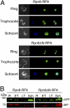
Intraerythrocytic expression of Rpn6 and Rpn6ΔN. (A) Stages of the erythrocytic life cycle of Rpn6-RFA (Upper) and Rpn6ΔN-RFA (Lower) parasites observed by live fluorescence microscopy. Images left to right are phase, DAPI, GFP, and fluorescence merge. (B) Western blot of lysates from different erythrocytic life cycle stages of Rpn6-RFA (Left) and Rpn6ΔN-RFA (Right) parasites, probed with anti-GFP antibody. BiP is the loading control. ET, early trophozoites; LT, late trophozoites; R, rings; Sch., schizonts.
Fig. 3.
Fig. 3.
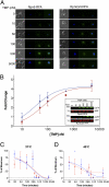
Regulation of RFA-tagged proteins. (A) Live fluorescence images of Rpn6-RFA (Left) and Rpn6ΔN-RFA (Right) parasites incubated for 24 h in different TMP doses (as indicated). Images left to right are phase, DAPI, GFP, and fluorescence merge. (B) Stabilization of Rpn6-RFA (red filled circle) and Rpn6ΔN-RFA (dark blue filled triangle) by TMP. Data are shown as fold change over the no-TMP sample. GFP signal from Western blots was normalized against BiP. (Inset) One of four independent TMP dose–response Western blots. BiP served as the loading control. (C and D) Change in the amount of protein over time when Rpn6-RFA (red filled circle) and Rpn6ΔN-RFA (dark blue filled triangle) parasites were incubated without TMP and quantified in three independent experiments. (Sample Western blots are shown in Fig. S5.) BiP served as the loading control. The experiment was done at two temperatures, 37 °C (C) and 40 °C (D). Error bars represent SE from three independent experiments.
Fig. 4.
Fig. 4.
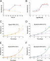
Rpn6 is an essential gene. (A) Synchronous ring-stage parasites were grown with (Rpn6-RFA, red filled circle; Rpn6ΔN-RFA. dark blue filled triangle) or without (Rpn6-RFA, orange filled diamond; Rpn6ΔN-RFA, light blue inverted filled triangle) 5 μM TMP, and their growth was monitored over 3 d by flow cytometry. (B) Asynchronous Rpn6-RFA (red filled circle) and Rpn6ΔN-RFA (dark blue filled triangle) parasites were incubated with different TMP concentrations, and their growth was measured after 48 h by flow cytometry. (C) Asynchronous Rpn6-RFA parasites were incubated with different TMP concentrations [5 μM (red filled circle); 0.312 μM (green filled square); 0.156 μM (orange filled diamond); 0.078 μM (open purple circle); or 0.039 μM (open yellow square)] at 37 °C, and their growth was monitored over 6 d by flow cytometry. (D) Asynchronous Rpn6ΔN-RFA parasites were incubated with different TMP concentrations [5 μM (dark blue filled triangle); 0.312 μM (light blue inverted filled triangle); 0.156 μM (inverted open green triangle); 0.078 μM (open purple triangle); and 0.039 μM (open purple diamond)] at 37 °C, and their growth was monitored over 6 d by flow cytometry. (E) Asynchronous Rpn6-RFA parasites were incubated with different TMP concentrations (symbols as in C) and were heat shocked for 6 h at 40 °C. After 6 h the cultures were transferred to 37 °C, and their growth was monitored over 6 d by flow cytometry. (F) Asynchronous Rpn6ΔN-RFA parasites were incubated with different TMP concentrations (symbols as in D) and were heat shocked for 6 h at 40 °C. After 6 h the cultures were transferred to 37 °C, and their growth was monitored over 6 d by flow cytometry. Error bars represent SE from one of three independent experiments done in triplicate.
Fig. 5.
Fig. 5.
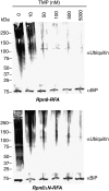
Ubiquitinated protein degradation depends on Rpn6. Synchronous early-trophozoite-stage Rpn6-RFA (Upper) and Rpn6ΔN-RFA (Lower) parasites were incubated for 24 h in different TMP concentrations (as indicated). Western blots of whole-cell protein extracts were probed with anti-ubiquitin antibody to assess the accumulation of ubiquitinated proteins in parasites. BiP was the loading control.

Similar articles

Cited by

References

    1. Breman JG. The ears of the hippopotamus: Manifestations, determinants, and estimates of the malaria burden. Am J Trop Med Hyg. 2001;64(Suppl1–2):1–11. - PubMed
    1. Singh GP, et al. Hyper-expansion of asparagines correlates with an abundance of proteins with prion-like domains in Plasmodium falciparum. Mol Biochem Parasitol. 2004;137:307–319. - PubMed
    1. Perutz MF, Pope BJ, Owen D, Wanker EE, Scherzinger E. Aggregation of proteins with expanded glutamine and alanine repeats of the glutamine-rich and asparagine-rich domains of Sup35 and of the amyloid beta-peptide of amyloid plaques. Proc Natl Acad Sci USA. 2002;99:5596–5600. - PMC - PubMed
    1. DePristo MA, Zilversmit MM, Hartl DL. On the abundance, amino acid composition, and evolutionary dynamics of low-complexity regions in proteins. Gene. 2006;378:19–30. - PubMed
    1. Zilversmit MM, et al. Low-complexity regions in Plasmodium falciparum: Missing links in the evolution of an extreme genome. Mol Biol Evol. 2010;27:2198–2209. - PMC - PubMed

Publication types

MeSH terms

LinkOut - more resources