. 2011 Mar 15;108(11):4376-81.
doi: 10.1073/pnas.1010647108. Epub 2011 Feb 28.

Butyrophilin-like 1 encodes an enterocyte protein that selectively regulates functional interactions with T lymphocytes

Affiliations

Butyrophilin-like 1 encodes an enterocyte protein that selectively regulates functional interactions with T lymphocytes

Anna Bas et al. Proc Natl Acad Sci U S A. .

Abstract

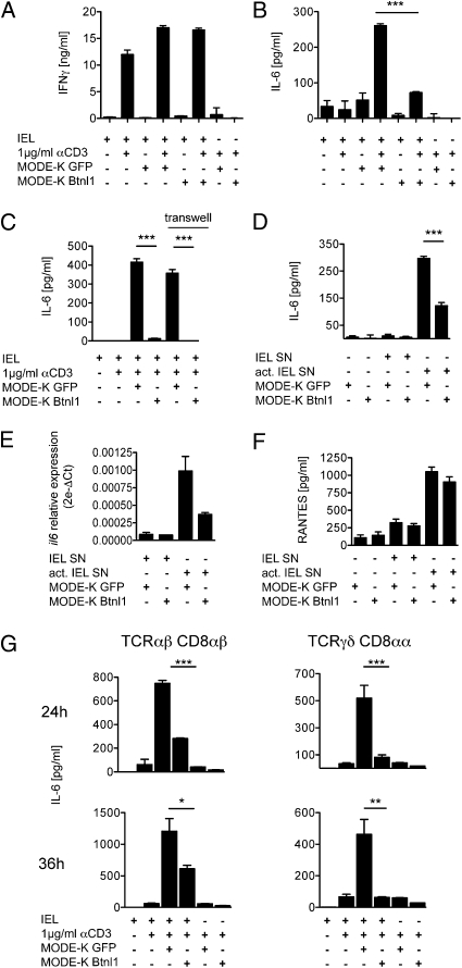
Although local regulation of T-cell responses by epithelial cells is increasingly viewed as important, few molecules mediating such regulation have been identified. Skint1, a recently identified member of the Ig-supergene family expressed by thymic epithelial cells and keratinocytes, specifies the murine epidermal intraepithelial lymphocyte (IEL) repertoire. Investigating whether Skint1-related molecules might regulate IEL in other compartments, this study focuses on buytrophilin-like 1 (Btnl1), which is conspicuously similar to Skint1 and primarily restricted to small intestinal epithelium. Btnl1 protein is mostly cytoplasmic, but surface expression can be induced, and in vivo Btnl1 can be detected adjacent to the IEL. In a newly developed culture system, enforced epithelial cell expression of Btnl1 attenuated the cells' response to activated IEL, as evidenced by suppression of IL-6 and other inflammatory mediators. These findings offer a unique perspective on emerging genetic data that Btnl genes may comprise novel and important local regulators of gut inflammation.


Conflict of interest statement

The authors declare no conflict of interest.

Figures

Fig. 1.
Fig. 1.
Structural analysis of Btnl1, -4, and -6. (A) Dot matrix comparison of Skint1 (y axis) with Btnl1, -4, and -6, and Skint2, and B7.1. (B) Full-length Btnl1, -4, and -6 cDNA and minor splice variants. Hatched boxes depict exons with internal in-frame deletions. The 5′ untranslated exon, U, identified for Btnl1 remains putative for Btnl4 and -6. SP, signal peptide; TM, transmembrane domain. (C) Inferred protein organization of Btnl1, -4, and -6, with predicted lengths of each domain as predicted by Pfam, SMART, TMHMM, and SignalP databases.
Fig. 2.
Fig. 2.
Quantitative RT-PCR analysis of Btnl1, -4, and -6 mRNA (A) in normal mouse tissues; (B) in CD45(−) small intestinal epithelial cells (IECs) versus CD45(+) small intestinal IEL; (C) in large intestines harvested from three mice; and (D) in the small intestines of two mice of each of three strains having low (Balb/CJ, DBA/2J, C57L/J) or high numbers of Vδ4(+) IEL (B10.BR, CBA/J, C58/J) (different symbols depict different strains). **P < 0.005 for comparison by unpaired two-tailed t test of means and data-points distribution. Error bars in B and C indicate SEM.
Fig. 3.
Fig. 3.
Btnl1 Protein. (A) A Btnl1-reactive antibody was raised against a synthetic peptide (residues 82–96); peptide alignment across Btnl1, -4, and -6 is shown. (B) Cleared lysates of enterocytes (lane 1) and liver (lane 2) resolved on nonreducing or reducing SDS/PAGE were Western-blotted with preimmune rabbit serum or peptide-purified anti-Btnl1 antibody. The GAPDH immunoblot acts as a loading control; size markers are in kilodaltons; specific antibody-reactive species are shown by arrows. (C) Btnl1 immunopurified from enterocyte lysates, was treated with N-glycosidase F (degly) as indicated, and the protein resolved as in B. (D) IECs derived from different mice at different times from different colonies, gated on CD45(−) cells (Upper), were stained with peptide-purified Btnl1-specific antibody (solid black line) or preimmune serum (shaded histogram). (E) Cultured enterocytes were treated with IFN-γ or activated IEL supernatant diluted 1:10 in enterocyte medium, or left untreated for 24 h, and then stained for Btnl1 and MHCII. Two independent experiments are shown. Differences in Btnl1 staining levels are partly because of different secondary reagents used, labeled with either AlexaFluor 488 or APC.
Fig. 4.
Fig. 4.
Localization of Btnl1 in murine small intestine. Small intestinal sections were immunostained with peptide-purified anti-Btnl1 antibody (red in B–D and G–I; brown in E and J), with antibody (anti-A33) to a well-established enterocyte surface marker (green in G) or with anti-CD3 (green in H and I; blue in J). Btnl1+ epithelial cells were detected in most villi, equally distributed from crypts to tips (B and C). Btnl1 adopted either a lobular/reticular pattern, exemplified by long arrows in D, or a lateral pattern, exemplified by arrowheads in D and arrows in E. Triple-staining for A33, Btnl1, and nuclei indicated that Btnl1 most commonly localized to a perinuclear region (G). Sections stained for Btnl1 and CD3 indicated prominent incidences of close juxtaposition, as depicted by arrows in H to J. No staining was detected using preimmune serum (A and F). Sections A to D and G and H were counterstained with DAPI (blue) to visualize nuclei.
Fig. 5.
Fig. 5.
Btnl1 selectively modulates cytokine expression. Culture of total IEL (A–C) or TCRγδ and TCRαβ IEL subsets (G) with (+) or without (−) anti-CD3, and with or without MODE-K cells transfected with gfp, or Btnl1-IRES-gfp, respectively, as indicated. In C, transwells separated IELs and MODE-K. Alternatively, MODE-K.gfp or MODE-K.gfp.Btnl1 cells were cultured with supernatants (SN) from activated or unactivated IEL (D–F). IFN-γ, IL-6, and RANTES were assessed by ELISA (A–D, F) and Luminex (G). Relative expression of il6 mRNA was determined by quantitative RT-PCR (E). *P < 0.05, **P ≤ 0.005, and ***P ≤ 0.0001 as calculated for means and SD by unpaired two-tailed t test.

References

    1. Kapsenberg ML. Dendritic-cell control of pathogen-driven T-cell polarization. Nat Rev Immunol. 2003;3:984–993. - PubMed
    1. Zaph C, et al. Epithelial-cell-intrinsic IKK-beta expression regulates intestinal immune homeostasis. Nature. 2007;446:552–556. - PubMed
    1. Swamy M, Jamora C, Havran W, Hayday A. Epithelial decision makers: In search of the “epimmunome”. Nat Immunol. 2010;11:656–665. - PMC - PubMed
    1. Hayday A, Theodoridis E, Ramsburg E, Shires J. Intraepithelial lymphocytes: Exploring the Third Way in immunology. Nat Immunol. 2001;2:997–1003. - PubMed
    1. Komano H, et al. Homeostatic regulation of intestinal epithelia by intraepithelial gamma delta T cells. Proc Natl Acad Sci USA. 1995;92:6147–6151. - PMC - PubMed

Publication types

MeSH terms