. 2011 Mar 15;108(11):4447-52.
doi: 10.1073/pnas.1101219108. Epub 2011 Feb 28.

Deficiency of Dgcr8, a gene disrupted by the 22q11.2 microdeletion, results in altered short-term plasticity in the prefrontal cortex

Affiliations

Deficiency of Dgcr8, a gene disrupted by the 22q11.2 microdeletion, results in altered short-term plasticity in the prefrontal cortex

Karine Fénelon et al. Proc Natl Acad Sci U S A. .

Abstract

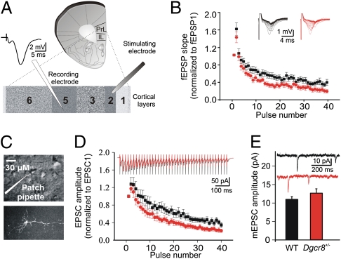
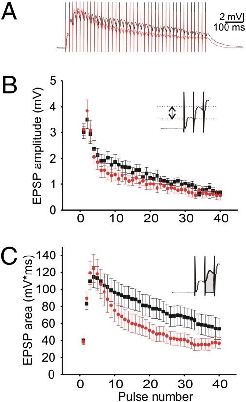
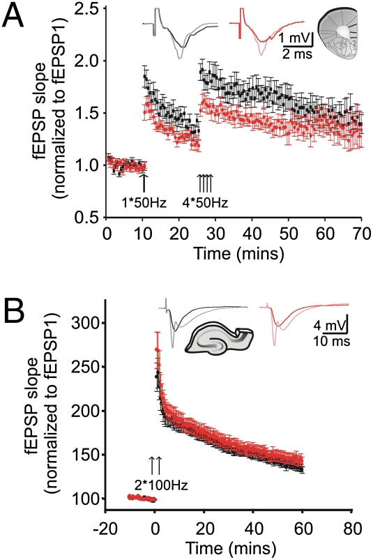
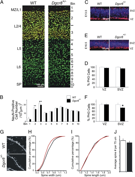
Individuals with 22q11.2 microdeletions have cognitive and behavioral impairments and the highest known genetic risk for developing schizophrenia. One gene disrupted by the 22q11.2 microdeletion is DGCR8, a component of the "microprocessor" complex that is essential for microRNA production, resulting in abnormal processing of specific brain miRNAs and working memory deficits. Here, we determine the effect of Dgcr8 deficiency on the structure and function of cortical circuits by assessing their laminar organization, as well as the neuronal morphology, and intrinsic and synaptic properties of layer 5 pyramidal neurons in the prefrontal cortex of Dgcr8(+/-) mutant mice. We found that heterozygous Dgcr8 mutant mice have slightly fewer cortical layer 2/4 neurons and that the basal dendrites of layer 5 pyramidal neurons have slightly smaller spines. In addition to the modest structural changes, field potential and whole-cell electrophysiological recordings performed in layer 5 of the prefrontal cortex revealed greater short-term synaptic depression during brief stimulation trains applied at 50 Hz to superficial cortical layers. This finding was accompanied by a decrease in the initial phase of synaptic potentiation. Our results identify altered short-term plasticity as a neural substrate underlying the cognitive dysfunction and the increased risk for schizophrenia associated with the 22q11.2 microdeletions.


Conflict of interest statement

The authors declare no conflict of interest.

Figures

Fig. 1.
Fig. 1.
The effect of the Dgcr8 deficiency on STD in L5 of mPFC. (A) Schematic representation of the prelimbic (PrL) and infralimbic (IL) areas in the mPFC slice preparation. The stimulating electrode was placed at the border of L1 and L2/3, and the extracellular recording electrode was placed in L5. A sample field fEPSP is shown. (B) Field EPSPs evoked by 40 stimuli at 50 Hz. The upper superimposed sample traces are individual fEPSP responses to each stimulus within the 50-Hz train. STD is significantly greater in Dgcr8+/− mice (N = 7; n = 16) than in the WT controls (N = 8; n = 14) (two-way repeated-measures ANOVA; P < 0.02). Post hoc analysis revealed that the level of depression was significantly greater in the Dgcr8+/− mice compared with WT mice at the seventh pulse (P = 0.048). (C) Voltage clamp recordings were made from visually identified (Upper) and biocytin-filled (Lower) L5 pyramidal neurons of the mPFC. (D) (Upper) Traces show the decrease in EPSC amplitudes in response to 40 pulses applied at 50 Hz and are the averages of the 10 repetitions performed in WT (N = 8, n = 13) and in Dgcr8+/− mice (N = 9, n = 11). Each vertical line represents the stimulation artifact. The short-term depression of the mean EPSC amplitudes is significantly greater in the mutant mice than in WT controls across experiments (two-way repeated-measures ANOVA; P < 0.05). Post hoc analysis revealed that the level of depression was significantly greater in the Dgcr8+/− mice compared with WT mice at the 14th pulse (P = 0.033). (E) The mean amplitude of mEPSCs was not different between genotypes. WT mice (N = 4; n = 8); Dgcr8+/− mice (N = 3; n = 4). Sample mEPSC traces of recordings from WT (black) and Dgcr8+/− (red) mice are shown.
Fig. 2.
Fig. 2.
Synaptic summation of EPSPs in L5 pyramidal neurons of the mPFC of Dgcr8+/− mice. (A) The traces are the averages of the 10 repetitions performed in WT (black; N = 10, n = 12) and Dgcr8+/− mice (red; N = 10, n = 15). Each vertical line is a stimulation artifact. (B) Across experiments, the mean amplitude of each individual EPSP measured from a baseline obtained just after each pulse during the 50-Hz train is unchanged by the mutation (two-way repeated-measures ANOVA; P > 0.05). (C) In contrast, the mean EPSP areas measured from the initial resting level and which includes the summating effect of the previous EPSPs in the train was significantly decreased by the mutation (WT mice; Dgcr8+/− mice; two-way repeated-measures ANOVA; P < 0.0001). The Insets in B and C show the procedures used for measuring the size of the EPSPs.
Fig. 3.
Fig. 3.
Synaptic plasticity in L5 of the mPFC and in CA1 of Dgcr8+/− mice. (A) Synaptic potentiation in WT mice (N = 8, n = 12) and Dgcr8+/− mice (N = 7, n = 14). There is a significant difference in the degree of potentiation of fEPSPs over time (two-way repeated-measures ANOVA; P < 0.05). Immediately after the first 50-Hz train (first arrow), the level of potentiation is significantly lower in the Dgcr8+/− mice. Similarly, after the four consecutive 50-Hz trains (four arrows), post hoc testing revealed that the difference in potentiation lasts for about 20 min. In contrast, long-term potentiation assayed 40 min after the tetanization is unaffected by the mutation. (Inset) Sample fEPSPs traces before (WT and Dgcr8+/− mice are black and red traces, respectively) and immediately after the first tetanization (WT and Dgcr8+/− are gray and pink traces, respectively). (B) Synaptic potentiation induced in CA1 by two trains (arrows) of 100 Hz (100 pulses, 1 s) applied to Schaffer collaterals is normal in Dgcr8+/− mice (two-way repeated-measures ANOVA; P > 0.05). WT mice (N = 5; n = 20); Dgcr8+/− mice (N = 5; n = 20). (Inset) Sample fEPSPs traces before (WT and Dgcr8+/− mice are black and red traces, respectively) and immediately after the tetanization (WT and Dgcr8+/− are gray and pink traces, respectively).
Fig. 4.
Fig. 4.
Structural changes in the cortex of Dgcr8+/− mice. (A) Schematic representation of probe locations for quantifying frequency of neurons labeled with a pan-neuronal marker (NeuN, green) and L2/4 selective marker (Cux1, red) in the cortex of 6-wk-old WT and Dgcr8+/− mice. (B) NeuN-labeled cells in medial frontal cortex of WT and Dgcr8+/− mice. The frequency of NeuN-labeled cells across 10 equal bins from pia to white matter at the medial region of the frontal cortex is shown: frequency of NeuN-labeled cells is represented as number of cells per 104 mm2. (C–F) Progenitor cells were counted throughout the entire cortex at E13.5 (C and D, n = 17 per genotype) and E16.5 (E and F, n = 16 per genotype) and normalized to corresponding WT values. Basal and apical progenitors, both identified by phosphohistone 3 (PH3) labeling (red), are distinguished by their positions in the SVZ versus VZ via immunostaining for Tbr-2 (blue), a SVZ marker. (G) High-magnification representative images of basal dendrites of EGFP-expressing pyramidal neurons in the L5 region of the mPFC of WT Thy1-GFP/M+/− (n = 5) and Dgcr8+/−;Thy1-GFP/M+/− (n = 5) mice. (H and I) Analysis of the width and length of mushroom spines in Dgcr8+/−;Thy1-GFP/M+/− L5 neurons basal dendrites. Reduction in the width (H), but not length (I) was observed. (J) The density of all spines (estimated over 75 μm of dendritic length) was unaffected in Dgcr8+/−;Thy1-GFP/M+/− neurons. Data are shown as mean ± SEM. *P < 0.05, **P < 0.001.

References

    1. Karayiorgou M, Simon TJ, Gogos JA. 22q11.2 microdeletions: Linking DNA structural variation to brain dysfunction and schizophrenia. Nat Rev Neurosci. 2010;11:402–416. - PMC - PubMed
    1. Karayiorgou M, et al. Schizophrenia susceptibility associated with interstitial deletions of chromosome 22q11. Proc Natl Acad Sci USA. 1995;92:7612–7616. - PMC - PubMed
    1. Stark KL, et al. Altered brain microRNA biogenesis contributes to phenotypic deficits in a 22q11-deletion mouse model. Nat Genet. 2008;40:751–760. - PubMed
    1. Tomari Y, Zamore PD. MicroRNA biogenesis: Drosha can't cut it without a partner. Curr Biol. 2005;15(2):R61–R64. - PubMed
    1. Elvevåg B, Goldberg TE. Cognitive impairment in schizophrenia is the core of the disorder. Crit Rev Neurobiol. 2000;14(1):1–21. - PubMed

Publication types

Supplementary concepts

LinkOut - more resources