. 2011 Mar 22;108(12):4852-7.
doi: 10.1073/pnas.1017667108. Epub 2011 Feb 28.

Cancer cell-derived microvesicles induce transformation by transferring tissue transglutaminase and fibronectin to recipient cells

Affiliations

Cancer cell-derived microvesicles induce transformation by transferring tissue transglutaminase and fibronectin to recipient cells

Marc A Antonyak et al. Proc Natl Acad Sci U S A. .

Erratum in

  • Proc Natl Acad Sci U S A. 2011 Oct 18;108(42):17569

Abstract

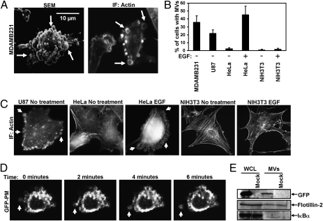
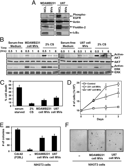
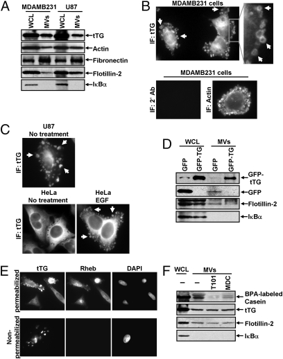
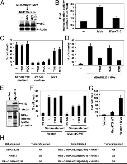
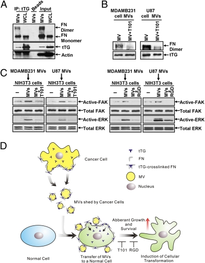
Tumor progression involves the ability of cancer cells to communicate with each other and with neighboring normal cells in their microenvironment. Microvesicles (MV) derived from human cancer cells have received a good deal of attention because of their ability to participate in the horizontal transfer of signaling proteins between cancer cells and to contribute to their invasive activity. Here we show that MV may play another important role in oncogenesis. In particular, we demonstrate that MV shed by two different human cancer cells, MDAMB231 breast carcinoma cells and U87 glioma cells, are capable of conferring onto normal fibroblasts and epithelial cells the transformed characteristics of cancer cells (e.g., anchorage-independent growth and enhanced survival capability) and that this effect requires the transfer of the protein cross-linking enzyme tissue transglutaminase (tTG). We further demonstrate that tTG is not sufficient to transform fibroblasts but rather that it must collaborate with another protein to mediate the transforming actions of the cancer cell-derived MV. Proteomic analyses of the MV derived from MDAMB231 and U87 cells indicated that both these vesicle preparations contained the tTG-binding partner and cross-inking substrate fibronectin (FN). Moreover, we found that tTG cross-links FN in MV from cancer cells and that the ensuing MV-mediated transfers of cross-linked FN and tTG to recipient fibroblasts function cooperatively to activate mitogenic signaling activities and to induce their transformation. These findings highlight a role for MV in the induction of cellular transformation and identify tTG and FN as essential participants in this process.


Conflict of interest statement

The authors declare no conflict of interest.

Figures

Fig. 1.
Fig. 1.
Distinct types of human cancer cells generate MV. (A) MDAMB231 cells were analyzed by SEM (Left) and immunofluorescent microscopy (IF) using rhodamine-conjugated phalloidin to detect F-actin (Right). Some of the largest MV are indicated by arrows. (B) Quantification of MV production by various cell lines cultured under serum-starved or EGF-stimulated conditions. Cells generating MV were detected by labeling the samples with rhodamine-conjugated phalloidin. The data shown represent the mean ± SD from three independent experiments. (C) Images of cells from the experiment performed in B. Some of the MV are denoted by arrows. (D) MDAMB231 cells transiently expressing a GFP-tagged form of the plasma membrane-targeting sequence (GFP-PM) from the Lyn tyrosine kinase were subjected to live-imaging fluorescent microscopy. Shown are a series of time-lapse images of a transfectant taken at 2-min intervals. The arrow indicates an MV that forms and is shed from a cell. (E) Serum-deprived MDAMB231 cells that were mock transfected or transfected with pEGFP were lysed, and the MV shed into the medium by the transfectants were isolated and lysed as well. The WCL and the MV lysates then were immunoblotted with antibodies against GFP, the MV marker flotillin-2, and the cytosolic-specific marker IκBα.
Fig. 2.
Fig. 2.
MDAMB231 and U87 cancer cell-derived MV are capable of transforming normal fibroblasts. (A) WCL of serum-starved MDAMB231 and U87 cells, as well as lysates of the MV shed by these cells, were immunoblotted with antibodies against the MV markers actin and flotillin-2, the cytosolic-specific marker IκBα, and the activated (phospho)-EGF receptor. (BD) Multiple sets of serum-deprived NIH 3T3 fibroblasts were incubated with serum-free medium, medium containing 2% CS, or medium supplemented with intact MV derived from either MDAMB231 or U87 cells as indicated. (B) One set of cells was lysed after being exposed to the various culturing conditions for the indicated lengths of time and then was immunoblotted with antibodies that recognize the activated and total forms of AKT and ERK. Two additional sets of fibroblasts were evaluated for their abilities to undergo serum deprivation-induced cell death (C) and to grow in low serum (2% CS) (D). For the growth assays, the culturing medium (including the MV) was replenished daily. (E) NIH 3T3 fibroblasts incubated with or without MV derived from MDAMB231 or U87 cells were subjected to anchorage-independent growth assays. The soft agar cultures were re-fed (including adding freshly prepared MV) every third day. NIH 3T3 cells expressing Cdc42 F28L were used as a positive control for these experiments. (F) Images of the resulting colonies that formed in E. The data shown in CE represent the mean ± SD from at least three independent experiments.
Fig. 3.
Fig. 3.
MV shed by cancer cells contain tTG. (A) WCL of serum-starved MDAMB231 and U87 cells, as well as lysates of the MV shed by these cells, were immunoblotted with several antibodies, including one against tTG. (B) (Upper Left) MDAMB231 cells immunostained with a tTG antibody. (Upper Right) The boxed area was enlarged, and arrows indicated certain MV. (Lower) An MDAMB231 cell costained with only the secondary antibody (Left) and with rhodamine-conjugated phalloidin to label the MV (Right). (C) Images of serum-starved U87 glioma cells and HeLa cervical carcinoma cells that were left untreated or were stimulated with EGF for 15 min as indicated and then were immunostained with a tTG antibody. Pronounced MV are indicated by arrows. (D) WCL of MDAMB231 cells ectopically expressing either GFP only or GFP-tTG, as well as lysates of the MV shed by these transfectants into their culturing medium, were immunoblotted with antibodies against GFP, the MV marker flotillin-2, and the cytosolic-specific marker IκBα. (E) Fluorescent images of permeabilized and nonpermeabilized samples of MDAMB231 cells stained with antibodies against tTG, the intracellular protein Ras homolog enriched in brain (Rheb), and DAPI to label nuclei. (F) WCL of serum-starved MDAMB231 cells and intact MV generated by these cells treated with or without the tTG inhibitors T101 (cell impermeable) or MDC (cell permeable), were assayed for transamidation activity as read out by the incorporation of BPA into casein. The samples then were immunoblotted with antibodies against tTG, flotillin-2, and IκBα.
Fig. 4.
Fig. 4.
The ability of MDAMB231 cell-derived MV to induce cellular transformation requires the transfer of tTG from MV to recipient cells. (A and B) Extracts of serum-starved NIH 3T3 fibroblasts that were incubated with serum-free medium or serum-free medium supplemented with MDAMB231 cell-derived MV that had been pretreated for 30 min with or without the tTG inhibitor T101 were immunoblotted with tTG and actin antibodies (A) and assayed for transamidation activity as read out by the incorporation of BPA into lysate proteins (B). (C) Cell death assays were performed on fibroblasts maintained in serum-free medium, 2% CS medium, or serum-free medium containing MDAMB231 cell-derived MV. Each culturing medium was left unsupplemented or was supplemented further with the tTG inhibitors T101 (cell impermeable) or MDC (cell permeable) as indicated. (D) Anchorage-independent growth assays were performed on fibroblasts incubated with MDAMB231 cell-derived MV that were untreated or treated with T101, the RGD peptide, or the control RGE peptide. (E) Lysates of NIH 3T3 cells stably overexpressing vector alone or Myc-tTG were immunoblotted with Myc and actin antibodies and were assayed for transamidation activity as read out by the incorporation of BPA into lysate proteins. (F) Cell death assays were performed on the NIH 3T3 stable cell lines maintained in serum-free medium treated with T101, MDC, or 2% CS or left untreated. (G) Anchorage-independent growth assays were performed on the NIH 3T3 stable cell lines. Vector-control fibroblasts were incubated with MDAMB231 cell-derived MV as a positive control. The data shown in BD, F, and G represent the mean ± SD from at least three independent experiments. (H) Tumor-formation assays were performed in which 5 × 10⁵ MDAMB231 cells mitotically arrested using mitomycin C (Mito-C-MDAMB231) expressing either control siRNA (siCont) or tTG siRNAs (siTG-1 or siTG-2) were injected s.c. alone or combined with 5 × 10⁵ NIH 3T3 fibroblasts into nude mice. As controls, untreated MDAMB231 and NIH 3T3 cells were injected into nude mice. The resulting tumors that formed for each condition were counted, and the results are shown in the table.
Fig. 5.
Fig. 5.
tTG cooperates functionally with FN to mediate the transforming actions of MV on recipient fibroblasts. (A) WCL of MDAMB231 and lysates of the MV shed by these cells were immunoblotted (Input) or were subjected to immunoprecipitation using a tTG antibody (IP: tTG) and then immunoblotted with FN, tTG, and actin antibodies. Note the detection of cross-linked FN in the MV lanes (FN dimer). (B) Intact MV collected from MDAMB231 or U87 cells were treated with T101 or were left untreated before being lysed. The MV extracts then were immunoblotted with FN and tTG antibodies. Note that the cross-linked forms of FN detected in the MV samples (FN dimer) were reduced significantly by T101 treatment. (C) Lysates of fibroblasts that were incubated with or without MV derived from MDAMB231 and U87 cells that had been pretreated or not with T101 were immunoblotted with antibodies against FAK and ERK or with antibodies that specifically recognize the activated forms of these protein kinases. (D) Diagram depicting how MV transform recipient cells. MV containing tTG and fibronectin are generated and released from the surfaces of human cancer cells. The MV then can be taken up by or can directly alter the microenvironment of neighboring normal cells, where the cotransfer of tTG and FN function cooperatively on the recipient cells to induce signaling events that promote cell survival and aberrant cell growth.

Similar articles

Cited by

References

    1. Al-Nedawi K, Meehan B, Rak J. Microvesicles: Messengers and mediators of tumor progression. Cell Cycle. 2009;8:2014–2018. - PubMed
    1. Cocucci E, Racchetti G, Meldolesi J. Shedding microvesicles: Artefacts no more. Trends Cell Biol. 2009;19:43–51. - PubMed
    1. Ratajczak J, Wysoczynski M, Hayek F, Janowska-Wieczorek A, Ratajczak MZ. Membrane-derived microvesicles: Important and underappreciated mediators of cell-to-cell communication. Leukemia. 2006;20:1487–1495. - PubMed
    1. Skog J, et al. Glioblastoma microvesicles transport RNA and proteins that promote tumour growth and provide diagnostic biomarkers. Nat Cell Biol. 2008;10:1470–1476. - PMC - PubMed
    1. Al-Nedawi K, et al. Intercellular transfer of the oncogenic receptor EGFRvIII by microvesicles derived from tumour cells. Nat Cell Biol. 2008;10:619–624. - PubMed

Publication types