. 2011 Mar 15;108(11):4334-9.
doi: 10.1073/pnas.1017150108. Epub 2011 Feb 28.

Active liquid-like behavior of nucleoli determines their size and shape in Xenopus laevis oocytes

Affiliations

Active liquid-like behavior of nucleoli determines their size and shape in Xenopus laevis oocytes

Clifford P Brangwynne et al. Proc Natl Acad Sci U S A. .

Abstract

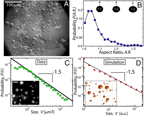
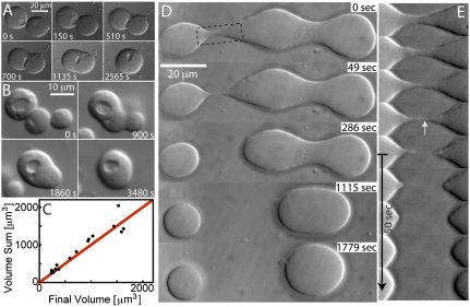
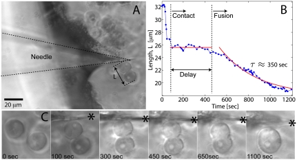
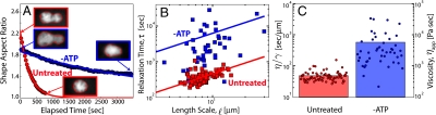
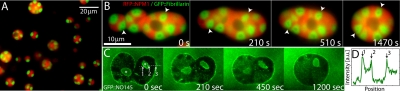
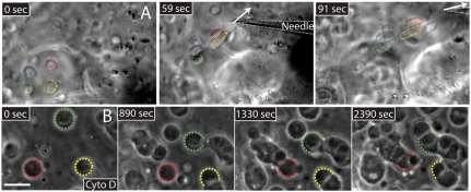
For most intracellular structures with larger than molecular dimensions, little is known about the connection between underlying molecular activities and higher order organization such as size and shape. Here, we show that both the size and shape of the amphibian oocyte nucleolus ultimately arise because nucleoli behave as liquid-like droplets of RNA and protein, exhibiting characteristic viscous fluid dynamics even on timescales of < 1 min. We use these dynamics to determine an apparent nucleolar viscosity, and we show that this viscosity is ATP-dependent, suggesting a role for active processes in fluidizing internal contents. Nucleolar surface tension and fluidity cause their restructuring into spherical droplets upon imposed mechanical deformations. Nucleoli exhibit a broad distribution of sizes with a characteristic power law, which we show is a consequence of spontaneous coalescence events. These results have implications for the function of nucleoli in ribosome subunit processing and provide a physical link between activity within a macromolecular assembly and its physical properties on larger length scales.


Conflict of interest statement

The authors declare no conflict of interest.

Figures

Fig. 1.
Fig. 1.
Size and shape of X. laevis oocyte nucleoli. (A) DIC image of nucleoli in the X. laevis GV. Nuclear bodies can be readily seen in DIC. Most of these are extrachromosomal nucleoli. (B) We plot the distribution of nucleolar aspect ratios obtained from analysis of GFP∷NPM1 images of nucleoli (Inset in C). The average is 1.07 ± 0.06; a perfect sphere would be 1.0. Thus, most nucleoli are highly spherical. (C) Distribution of nucleolar volume exhibits a power-law distribution with an exponent of -1.5. Inset shows an image of GFP∷NPM1–labeled nucleoli (scale bar, 20 μm). (D) Monte Carlo simulation of fusing droplets, with a slow constant influx of small droplets. The distribution of droplet volume exhibits power-law behavior with an exponent of -1.5. A snapshot of a subregion of the simulation is shown in the Inset.
Fig. 2.
Fig. 2.
Fluid-like behavior of nucleoli. (A) DIC image sequence showing fusion of two spherical nucleoli into one larger spherical nucleolus. (B) DIC image of the fusion of three spherical nucleoli into one larger spherical nucleolus. (C) Plot of the sum of nucleoli volumes before and after fusion. The red line corresponds to conserved volume. (D) In the first frame, three nucleoli that have come into contact and begun fusing are visible. The bridge between the two on the Left is unstable and pinches off, whereas the bridge between the nucleoli on the Right is stable and they fuse. (E) Close-up of the rupturing bridge from D, showing that the threads of nucleolar material are resorbed within 1 min after rupturing.
Fig. 3.
Fig. 3.
Needle-induced fusion of two nucleoli. (A) Image showing a microneedle that has been used to move two nucleoli together. (B) Time course of the distance between the two nucleoli. Initially they are not touching. At approximately 75 s, they are brought into contact with one another, but they do not appear to begin fusing until approximately 500 s, when they fuse into a single round sphere on a timescale of τ ∼ 350 s. (C) Images showing the sequence of events. The needle is visible in the second frame (needle tip position denoted by the asterisk).
Fig. 4.
Fig. 4.
Analysis of fusion dynamics and ATP dependence. (A) The red curve shows the dynamics of a single example control fusion event of two GFP∷NPM1–labeled nucleoli, shown by the images in the red box. The blue curve shows the slower fusion dynamics of a comparable pair of GFP∷NPM1 nucleoli (blue boxes) in an ATP-depleted GV. The red outline in the images is the output of the image analysis routines used to calculate aspect ratio. (B) Plot of the fusion time, τ, vs. length, , from untreated nucleoli (N = 77), and apyrase-treated nucleoli (N = 46). The red and blue lines are linear fits to the control and apyrase data, respectively. (C) Scatter plot of η/γ obtained from control and ATP-depleted nucleoli. The Right axis reflects the apparent viscosity, ηapp, obtained assuming a surface tension of γ = 10 μN/m.
Fig. 5.
Fig. 5.
Dynamics of nucleolar substructures. (A) A GV exhibiting GFP∷fibrillarin/RFP∷NPM1–labeled nucleoli. Note that larger nucleoli contain many fibrillarin cores, whereas smaller nucleoli contain only one or two fibrillarin cores. (B) Image sequence showing fusion of several GFP∷fibrillarin/RFP∷NPM1-labeled nucleoli. Arrowheads track the positions of two fibrillar cores. (C) Image sequence showing the dynamics of GFP∷NO145 at the cortex of two fusing nucleoli. Note the GFP∷NO145 localization to the interior of nucleoli at the surface of nucleolar vacuole-like structures (*). (D) Intensity profile across the dotted line shown in C; peaks 1 and 2 reflect vacuolar surface, and peak 3 reflects nucleolar cortex.
Fig. 6.
Fig. 6.
The role of a surrounding actin scaffold in slowing internucleolar contact. (A) Pulling on one region of the GV with a microneedle displaces nucleoli in distal regions, suggesting the presence of an elastic scaffold. Four nucleoli are highlighted. (B) GV treated with Cyto-D to disrupt filamentous actin. Nucleoli freely diffuse throughout the GV and fuse with one another upon contact; the fates of three nucleoli are highlighted. One large nucleolar clump results; note, however, that under these drug conditions nucleoli do not appear to regain sphericity and fusion appears perturbed.

Similar articles

Cited by

References

    1. Savas JN, et al. Huntington's disease protein contributes to RNA-mediated gene silencing through association with Argonaute and P bodies. Proc Natl Acad Sci USA. 2008;105:10820–10825. - PMC - PubMed
    1. Gall JG. Cajal bodies: The first 100 years. Annu Rev Cell Dev Biol. 2000;16:273–300. - PubMed
    1. Spector DL. Nuclear domains. J Cell Sci. 2001;114:2891–2893. - PubMed
    1. Brangwynne CP, et al. Germline P granules are liquid droplets that localize by controlled dissolution/condensation. Science. 2009;324:1729–1732. - PubMed
    1. Helpap B. Observations on the number, size and localization of nucleoli in hyperplastic and neoplastic prostatic disease. Histopathology. 1988;13:203–211. - PubMed

Publication types