. 2011 Mar 2;3(72):72ra17.
doi: 10.1126/scitranslmed.3001498.

Therapeutic targeting of SPINK1-positive prostate cancer

Affiliations

Therapeutic targeting of SPINK1-positive prostate cancer

Bushra Ateeq et al. Sci Transl Med. .

Abstract

Gene fusions involving ETS (erythroblastosis virus E26 transformation-specific) family transcription factors are found in ~50% of prostate cancers and as such can be used as a basis for the molecular subclassification of prostate cancer. Previously, we showed that marked overexpression of SPINK1 (serine peptidase inhibitor, Kazal type 1), which encodes a secreted serine protease inhibitor, defines an aggressive molecular subtype of ETS fusion-negative prostate cancers (SPINK1+/ETS⁻, ~10% of all prostate cancers). Here, we examined the potential of SPINK1 as an extracellular therapeutic target in prostate cancer. Recombinant SPINK1 protein (rSPINK1) stimulated cell proliferation in benign RWPE as well as cancerous prostate cells. Indeed, RWPE cells treated with either rSPINK1 or conditioned medium from 22RV1 prostate cancer cells (SPINK1+/ETS⁻) significantly increased cell invasion and intravasation when compared with untreated cells. In contrast, knockdown of SPINK1 in 22RV1 cells inhibited cell proliferation, cell invasion, and tumor growth in xenograft assays. 22RV1 cell proliferation, invasion, and intravasation were attenuated by a monoclonal antibody (mAb) to SPINK1 as well. We also demonstrated that SPINK1 partially mediated its neoplastic effects through interaction with the epidermal growth factor receptor (EGFR). Administration of antibodies to SPINK1 or EGFR (cetuximab) in mice bearing 22RV1 xenografts attenuated tumor growth by more than 60 and 40%, respectively, or ~75% when combined, without affecting PC3 xenograft (SPINK1⁻/ETS⁻) growth. Thus, this study suggests that SPINK1 may be a therapeutic target in a subset of patients with SPINK1+/ETS⁻ prostate cancer. Our results provide a rationale for both the development of humanized mAbs to SPINK1 and evaluation of EGFR inhibition in SPINK1+/ETS⁻ prostate cancers.


Figures
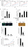
Figure 1
Figure 1
Oncogenic in vitro effects of SPINK1 in prostate cells. (A) SPINK1 stimulates cell proliferation in SPINK1-/ETS- cell lines. Benign immortalized prostate cell line RWPE and prostate cancer cell lines DU145 and PC3 (all SPINK1-/ETS-) were untreated or treated with 10ng/ml of rSPINK1. Cell proliferation was measured by a WST-1 colorimetric assay at the indicated time points. (B) SPINK1 mediates invasion of RWPE cells as measured by Boyden chamber Matrigel invasion assay. RWPE cells were treated with 10ng/ml of rSPINK1 or conditioned media (CM) from 22RV1 cells (SPINK1+/ETS-). (C) As in B, except using 22RV1 cells transfected with siRNA against SPINK1. SPINK1 silenced 22RV1 cells were further treated with 10ng/ml of rSPINK1 or CM from 22RV1 cells. (D) SPINK1 expression in SPINK1 knockdown 22RV1 cells (stable pooled shSPINK1 or stable shSPINK1 clone 11) compared to non-targeting pooled stable control (shNS vector) cells by quantitative PCR (transcript) or immunofluorescence using an antibody against SPINK1 (protein, upper inset; 600X magnification). (E) Invasion assay using shSPINK1 and shNS cells. Representative photomicrographs (400X magnification) showing cell motility assay (top inset) are shown. shNS vector cells exhibit longer cell motility tracks as compared to shSPINK1 knockdown cells. (F) Cell proliferation assay using pooled shSPINK1, shSPINK1 clone 11, or shNS cells at the indicated time points. (G) Soft agar colony assay using pooled shSPINK1 cells and shNS cells. All experiments were independently performed in triplicate. Data shown represents mean +/- SEM. P values from significant two-sided Student's t tests are given (* = <0.05, ** = <0.001).
Figure 2
Figure 2
Anti-SPINK1 mAb attenuates in vitro proliferation and invasion exclusively in SPINK1+/ETS- prostate cancer cells. (A) Cell proliferation of DU145, PC3 and 22RV1 cells was assessed in the presence of 1μg/ml SPINK1 mAb or IgG mAb (B) As in A except using 22RV1 cells and 0.5-1μg/ml SPINK1 mAb or IgG mAb. (C) Effect of SPINK1 mAb or IgG mAb on invasion of SPINK1+/ETS- cells (22RV1 and CWR22PC) and SPINK1+/ETS- cells (DU145, PC3, LNCaP and VCaP) All experiments were independently performed in triplicates. Data shown represents mean +/- SEM. P values from significant two-sided Student's t tests are given (* = <0.05, ** = <0.001).
Figure 3
Figure 3
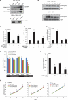
SPINK1 mediates its oncogenic effects in part through EGFR (A) Immunoprecipitation using anti-IgG, anti-SPINK1 or anti-GST of exogenous SPINK1-GST, GST or GST-VEGFR added to HEK-293 cells transfected with EGFR and immunoblotted with anti-EGFR (top panel), and immunoprecipitation using anti-IgG or anti-SPINK1 of exogenous SPINK1-GST added to 22RV1 cells and immunoblotted with anti-EGFR (bottom panel) (B) Western blot showing EGFR phosphorylation in response to rSPINK1 (100 ng/ml) or EGF (10 ng/ml) stimulation. (C) Invasion assay showing siRNA mediated EGFR knockdown 22RV1 cells treated with 10 ng/ml of rSPINK1 (D) Same as in C, except with RWPE cells. (E) Invasion assay showing rSPINK1 (10 ng/ml) stimulated RWPE cells in the presence or absence of C225 (cetuximab, 50 μg/ml) or IgG mAb (50 μg/ml) (F) Invasion assay showing the effect of IgG or C225 antibody on SPINK1+ and SPINK1- cancer cells. (G) As in F, except 22RV1 cells were treated with a combination of anti-SPINK1 and/or C225 mAb (1 μg/ml and 50 μg/ml respectively). (H) Cell proliferation assay using the indicated cells in the presence of IgG mAb or C225. All experiments were independently performed in triplicates. Data shown represents mean +/- SEM. P values from significant two-sided Student's t tests are given (* = <0.05, ** = <0.001).
Figure 4
Figure 4
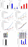
SPINK1 as a therapeutic target in SPINK1+ prostate cancer. (A) Chick chorioallantoic membrane (CAM) assay quantifying intravasated RWPE cells upon stimulation with rSPINK1 (n=6 in each group). (B) CAM assay using 22RV1 cells in the presence of IgG mAb, SPINK1 mAb or C225 (n=5 in each group), with fold change of intravasated cells compared to IgG mAb plotted. (C) As in B, except using PC3 cells. (D) Subcutaneous xenograft growth of shNS-luciferase (luc) or shSPINK1-luc 22RV1 cells implanted in male BALB/C nu/nu mice (n=10 in each group). (E) As in D, except using 22RV1-luc cell xenografts treated with control IgG mAb (n=8), SPINK1 mAb (n=6) or C225 (n=8) (10 mg/kg body weight) twice a week. (F) Same as in E, except mice (n=7 per group) were treated with a combination of SPINK1 mAb and C225 mAb (10 mg/kg body weight for both). (G) As in E & F, except using PC3-luc xenografts treated with control IgG mAb, SPINK1 mAb or C225 (n=8 per group) (10mg/kg body weight) alone or in combination twice a week. (H) Representative bioluminescence images from mice in D bearing pooled shNS-luc or shSPINK1-luc xenografts and % reduction in tumor volume at week 5. (I) Same as H, except bioluminescence images from mice bearing 22RV1-luc xenografts in (red, top panel) or PC3-luc (blue, lower panel) mice treated with IgG mAb, SPINK1 mAb, or C225 mAb alone or in combination, with comparative % reduction plot in tumor volume at week 5. Data shown represents mean +/- SEM. P values from significant two-sided Student's t tests are given (* = <0.05, ** = <0.001).

Comment in

Similar articles

Cited by

References

    1. Jemal A, Siegel R, Xu J, Ward E. Cancer statistics, 2010. CA Cancer J Clin. 2010;60:277–300. - PubMed
    1. Helgeson BE, Tomlins SA, Shah N, Laxman B, Cao Q, Prensner JR, Cao X, Singla N, Montie JE, Varambally S, Mehra R, Chinnaiyan AM. Characterization of TMPRSS2:ETV5 and SLC45A3:ETV5 gene fusions in prostate cancer. Cancer Res. 2008;68:73–80. - PubMed
    1. Tomlins SA, Rhodes DR, Perner S, Dhanasekaran SM, Mehra R, Sun XW, Varambally S, Cao X, Tchinda J, Kuefer R, Lee C, Montie JE, Shah RB, Pienta KJ, Rubin MA, Chinnaiyan AM. Recurrent fusion of TMPRSS2 and ETS transcription factor genes in prostate cancer. Science. 2005;310:644–648. - PubMed
    1. Tomlins SA, Mehra R, Rhodes DR, Smith LR, Roulston D, Helgeson BE, Cao X, Wei JT, Rubin MA, Shah RB, Chinnaiyan AM. TMPRSS2:ETV4 gene fusions define a third molecular subtype of prostate cancer. Cancer Res. 2006;66:3396–3400. - PubMed
    1. Tomlins SA, Laxman B, Dhanasekaran SM, Helgeson BE, Cao X, Morris DS, Menon A, Jing X, Cao Q, Han B, Yu J, Wang L, Montie JE, Rubin MA, Pienta KJ, Roulston D, Shah RB, Varambally S, Mehra R, Chinnaiyan AM. Distinct classes of chromosomal rearrangements create oncogenic ETS gene fusions in prostate cancer. Nature. 2007;448:595–599. - PubMed

Publication types

MeSH terms