Comparative Study
. 2011 Apr 1;186(7):4008-18.
doi: 10.4049/jimmunol.1003757. Epub 2011 Mar 2.

Simian immunodeficiency virus infection in the brain and lung leads to differential type I IFN signaling during acute infection

Affiliations
Comparative Study

Simian immunodeficiency virus infection in the brain and lung leads to differential type I IFN signaling during acute infection

Luna Alammar et al. J Immunol. .

Abstract

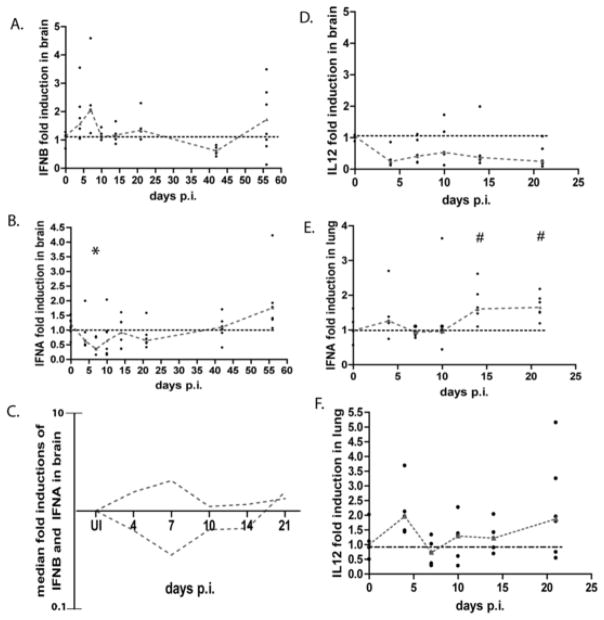
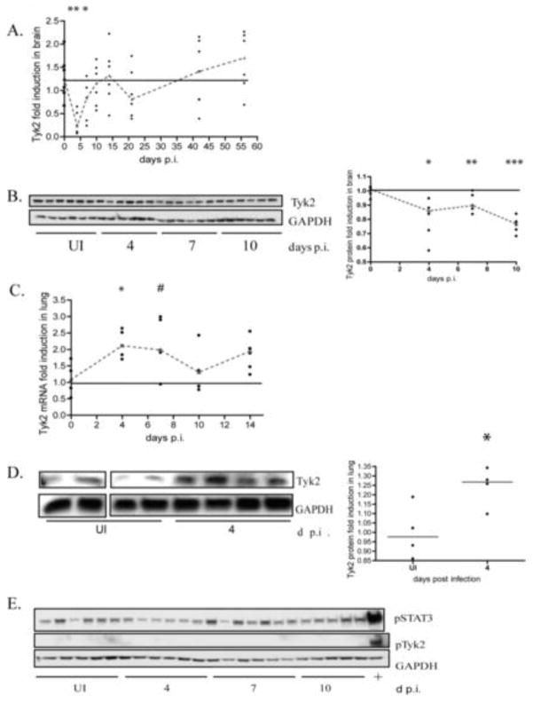
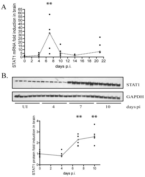
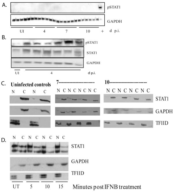
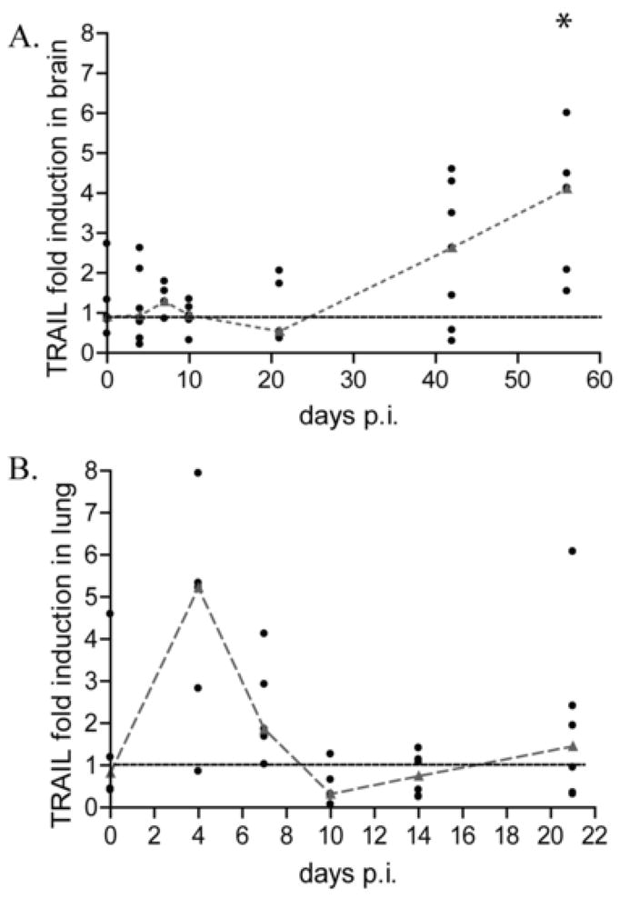
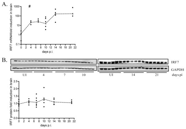
Using an accelerated and consistent SIV pigtailed macaque model of HIV-associated neurologic disorders, we have demonstrated that virus enters the brain during acute infection. However, neurologic symptoms do not manifest until late stages of infection, suggesting that immunological mechanisms exist within the CNS that control viral replication and associated inflammation. We have shown that IFN-β, a type I IFN central to viral innate immunity, is a major cytokine present in the brain during acute infection and is responsible for limiting virus infection and inflammatory cytokine expression. However, the induction and role of IFN-α in the CNS during acute SIV infection has never been examined in this model. In the classical model of IFN signaling, IFN-β signals through the IFN-α/β receptor, leading to expression of IFN-α. Surprisingly, although IFN-β is upregulated during acute SIV infection, we found that IFN-α is downregulated. We demonstrate that this downregulation is coupled with a suppression of signaling molecules downstream of the IFN receptor, namely tyrosine kinase 2, STAT1, and IFN regulatory factor 7, as indicated by either lack of protein phosphorylation, lack of nuclear accumulation, or transcriptional and/or translational repression. In contrast to brain, IFN-α is upregulated in lung and accompanied by activation of tyrosine kinase 2 and STAT1. These data provide a novel observation that during acute SIV infection in the brain, there is differential signaling through the IFN-α/β receptor that fails to activate expression of IFN-α in the brain.


Figures

Figure 1
Figure 1
Interferon alpha and IL-12 mRNA are down regulated in the brain, but up regulated in the lung during acute SIV infection. RNA was isolated from the brain and lung from SIV infected pigtailed macaques that were sacrificed at days 4, 7, 10, 14, 21, 42, and 56 as well as 6 uninfected controls and analyzed by qRT-PCR. Each animal is represented by a black dot, and dashes connect medians. Values are normalized to GAPDH (for alpha) and 18S ribosomal RNA (for IL-12) and are expressed as a ratio over the average of the uninfected controls (fold induction) using the ΔΔCt method (14). A. interferon beta mRNA expression in the brain. B and E, interferon alpha mRNA expression in brain and lung (respectively) * p=0.0101, # p=0.0519. Primers were made against conserved regions between all subtypes. C, overlay of the medians for mRNA expression of interferon beta and interferon alpha during SIV infection in the brain. D and F, IL-12 RNA expression in brain and lung, respectively.
Figure 2
Figure 2
Tyk2 mRNA and protein expression is down regulated during acute SIV infection in brain. A. qRT-PCR analysis for tyk2 on RNA isolated from the brain of pigtailed macaques sacrificed between days 4 and 56 p.i. including uninfected controls. 5–6 animals used per group. Values are normalized to 18S ribosomal RNA and expressed as fold induction. Medians are connected by red dashes. * p=0.0649, ** p= 0.0022. B. Western blot analysis of tyk2 protein in lysates from brain of pigtailed macaques sacrificed between days 4 and 10 p.i. including uninfected controls. 6 animals were used per group. All are normalized to GAPDH and expressed as fold induction over uninfected. Medians are connected by red dashes. * p=0.0087, **p=0.0152, *** p=0.0022 C. qRT-PCR analysis for tyk2 on RNA isolated from the lungs of the same pigtailed macaques sacrificed between days 4 and 14 p.i including uninfected controls. Values normalized to 18S ribosomal RNA. Medians are connected by red dashes. *p=0.0317, # p=0.0635. D. Western blot analysis of tyk2 protein on lysates made from lung of pigtailed macaques sacrificed on day 4 p.i including uninfected controls. Values normalized to GAPDH and expressed as fold induction. Medians are indicated by red dashes. * p=0.0571. E. Western blot analysis of ptyk2 (Y1054/1055) and P-STAT3 (S727) on the same protein lysates, as well as a lysate made from pigtailed macrophages stimulated with interferon beta (100 units/mL) for 10 minutes (designated “+”).
Figure 3
Figure 3
STAT1 mRNA and protein expression is up regulated during acute SIV infection. A. qRT-PCR analysis for STAT1 was done on RNA isolated from brain of pigtailed macaques sacrificed on days 4–21 p.i including uninfected controls. 6 animals were used for each group. Values are expressed as a fold induction normalized to 18S ribosomal RNA. Medians are indicated by red dashes. **p=0.0079 B. Western blot analysis for STAT1 done on protein lysates made from brain of macaques sacrificed on days 4–10 p.i. including uninfected controls (6 animals per group). Values are expressed as fold induction normalized to GAPDH. Medians are indicated by red dashes. **p=0.0022
Figure 4
Figure 4
Lack of STAT1 phosphorylation and nuclear accumulation in the brain during acute infection. A, western blot analysis of pSTAT1 (Y701) on the brain of pigtailed macaques as well as a lysate made from interferon beta stimulated macrophages harvested after 10 minutes (last sample designated “+”). B. western blot analysis of STAT1 and pSTAT1 (Y701) in lung protein lysates made from pigtailed macaques (5 animals) sacrificed at 4 days p.i and 1 uninfected control. C. nuclear (N) and cytoplasmic (C) lysates were made from the brain from animals sacrificed at 7 and 10 days p.i (4 each) as well as 2 uninfected controls. Western blot analysis for STAT1, GAPDH and TFIID were done on lysates. D. nuclear (N) and cytoplasmic (C) lysates were made from pigtailed macaque macrophages that were either untreated or treated with interferon beta for 5, 10 and 15 minutes. Western blot analysis for STAT1, GAPDH, and TFIID were done on lysates.
Figure 5
Figure 5
TRAIL mRNA expression is not induced in brain of pigtailed macaques during acute SIV infection. A, qRT-PCR analysis for TRAIL done on RNA isolated from brain from pigtail macaques sacrificed between days 4 and 56 p.i including uninfected controls (6 animals per group). * p=0.0157. B, qRT-PCR analysis for TRAIL done on RNA from lung isolated macaques sacrificed between days 4 and 21 days p.i. Values are normalized to 18S ribosomal RNA and represented as fold inductions over uninfected controls. Medians are connected by red dashes.
Figure 6
Figure 6
There is a post transcriptional block in IRF7 expression during acute SIV infection. A, qRT-PCR analysis for IRF7 on RNA isolated from brain from pigtailed macaques sacrificed between days 4 and 21 p.i. including uninfected controls (6 animals per group). Values are normalized to 18S ribosomal RNA and expressed as fold induction. Medians are connected by red dashes. # p=0.01. B, Protein lysates were made from brain of the same animals and western blot analysis was done for IRF7. Values are normalized to 18S ribosomal RNA and expressed as a fold induction over uninfected animals run on the same blot as the sample. Medians are indicated by red dashes.

Similar articles

Cited by

References

    1. Maschke M, Kastrup O, Esser S, Ross B, Hengge U, Hufnagel A. Incidence and prevalence of neurological disorders associated with HIV since the introduction of highly active antiretroviral therapy (HAART) J Neurol Neurosurg Psychiatry. 2000;69:376–380. - PMC - PubMed
    1. McArthur JC, Haughey N, Gartner S, Conant K, Pardo C, Nath A, Sacktor N. Human immunodeficiency virus-associated dementia: an evolving disease. J Neurovirol. 2003;9:205–221. - PubMed
    1. Sacktor N, McDermott MP, Marder K, Schifitto G, Selnes OA, McArthur JC, Stern Y, Albert S, Palumbo D, Kieburtz K, De Marcaida JA, Cohen B, Epstein L. HIV-associated cognitive impairment before and after the advent of combination therapy. J Neurovirol. 2002;8:136–142. - PubMed
    1. Fischer-Smith T, Rappaport J. Evolving paradigms in the pathogenesis of HIV-1-associated dementia. Expert Rev Mol Med. 2005;7:1–26. - PubMed
    1. Marra CM, Zhao Y, Clifford DB, Letendre S, Evans S, Henry K, Ellis RJ, Rodriguez B, Coombs RW, Schifitto G, McArthur JC, Robertson K. Impact of combination antiretroviral therapy on cerebrospinal fluid HIV RNA and neurocognitive performance. AIDS. 2009;23:1359–1366. - PMC - PubMed

Publication types

MeSH terms