Comparative Study
. 2011 Apr 5;76(14):1229-37.
doi: 10.1212/WNL.0b013e318214332c. Epub 2011 Mar 2.

Neuromyelitis optica unique area postrema lesions: nausea, vomiting, and pathogenic implications

Affiliations
Comparative Study

Neuromyelitis optica unique area postrema lesions: nausea, vomiting, and pathogenic implications

B F Gh Popescu et al. Neurology. .

Abstract

Objective: To characterize the neuropathologic features of neuromyelitis optica (NMO) at the medullary floor of the fourth ventricle and area postrema. Aquaporin-4 (AQP4) autoimmunity targets this region, resulting in intractable nausea associated with vomiting or hiccups in NMO.

Methods: This neuropathologic study was performed on archival brainstem tissue from 15 patients with NMO, 5 patients with multiple sclerosis (MS), and 8 neurologically normal subjects. Logistic regression was used to evaluate whether the presence of lesions at this level increased the odds of a patient with NMO having an episode of nausea/vomiting.

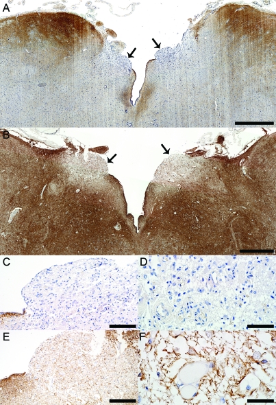
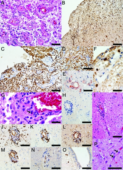
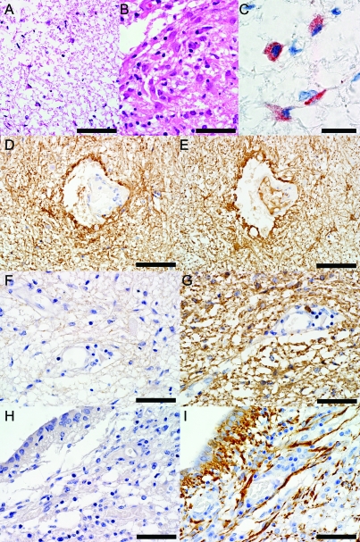
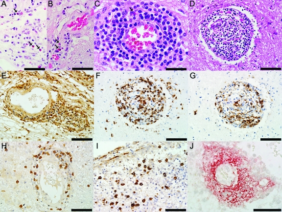
Results: Six patients with NMO (40%), but no patients with MS or normal controls, exhibited unilateral or bilateral lesions involving the area postrema and the medullary floor of the fourth ventricle. These lesions were characterized by tissue rarefaction, blood vessel thickening, no obvious neuronal or axonal pathology, and preservation of myelin in the subependymal medullary tegmentum. AQP4 immunoreactivity was lost or markedly reduced in all 6 cases, with moderate to marked perivascular and parenchymal lymphocytic inflammatory infiltrates, prominent microglial activation, and in 3 cases, eosinophils. Complement deposition in astrocytes, macrophages, and/or perivascularly, and a prominent astroglial reaction were also present. The odds of nausea/vomiting being documented clinically was 16-fold greater in NMO cases with area postrema lesions (95% confidence interval 1.43-437, p = 0.02).

Conclusions: These neuropathologic findings suggest the area postrema may be a selective target of the disease process in NMO, and are compatible with clinical reports of nausea and vomiting preceding episodes of optic neuritis and transverse myelitis or being the heralding symptom of NMO.


Figures

Figure 1
Figure 1. Aquaporin-4 (AQP4) is selectively lost in area postrema in neuromyelitis optica (NMO)
(A) The area postrema (arrows) in NMO patient 1 (patient 1 in table 1 and table e-1) who presented a clinically documented episode of nausea and intractable vomiting shows a characteristic selective loss of AQP4 (AQP4, scale bar = 1 mm). (B) In comparison, AQP4 is preserved in the area postrema (arrows) from a control patient with acute multiple sclerosis (Ctrl-MS) (AQP4, scale bar = 1 mm). (C) Higher magnification of the NMO area postrema (right arrow in A) shows a selective loss of AQP4, while subependymal immunoreactivity of AQP4 is preserved (AQP4, scale bar = 100 μm). (D) Higher magnification of the NMO area postrema (right arrow in A) shows the loss of the characteristic rim/rosette AQP4 distribution around blood vessels (AQP4, scale bar = 50 μm). (E) Higher magnification of area postrema (left arrow in B) shows preservation of AQP4 in Ctrl-MS (AQP4, scale bar = 200 μm). (F) Higher magnification of Ctrl-MS area postrema (left arrow in B) shows the preservation of AQP4 rim distribution around blood vessels (AQP4, scale bar = 50 μm).
Figure 2
Figure 2. Neuropathologic findings in the medullary floor of the fourth ventricle and area postrema of the neuromyelitis optica (NMO) patient 1 illustrated in figure 1, A, C, and D
(A) Thickening of the blood vessels in area postrema (hematoxylin & eosin [H&E], scale bar = 50 μm). (B) Profound activation of macrophages/microglia (KiM1P, scale bar = 100 μm). (C) Glial fibrillary acidic protein (GFAP) immunoreactive astrocytes extending foot processes that surrounded the endothelial cells are still present throughout these lesions (GFAP, scale bar = 50 μm). (D) GFAP immunoreactive astrocytes and preservation of neurons in the NMO area postrema (GFAP, scale bar = 50 μm). (E) Complement deposition in perivascular macrophages and astrocytes (C9neo, scale bar = 50 μm). (F) CD3+ T lymphocytes are present in the subependyma of the floor of the fourth ventricle (CD3, scale bar = 20 μm). (G) Eosinophils are components of the inflammatory infiltrates (H&E, scale bar = 20 μm). (H) Moderate perivascular inflammation in an area of aquaporin-4 (AQP4) loss (AQP4, scale bar = 50 μm). (I) Neurons are preserved around the blood vessel shown in H (H&E, scale bar = 50 μm). (J) Macrophages/microglia (KiM1P, scale bar = 50 μm), (K) CD3+ T lymphocytes (CD3, scale bar = 50 μm), (L) CD8+ T lymphocytes (CD8, scale bar = 50 μm), (M) B lymphocytes (CD20, scale bar = 50 μm), (N) Plasma cells (CD138, scale bar = 50 μm) are components of the perivascular inflammatory infiltrates. (O) Parenchymal plasma cells located in the subependyma of the floor of the fourth ventricle (CD138, scale bar = 50 μm). (P) Uninucleated, small, reactive astrocytes with thin, elongated processes (H&E, scale bar = 50 μm).
Figure 3
Figure 3. Astrocytic changes in neuromyelitis optica (NMO) area postrema lesions
(A) Astrogliosis in an NMO area of aquaporin-4 (AQP4) loss and tissue rarefaction in the ventricular floor with uninucleated small reactive astrocytes with elongated, thin processes (hematoxylin & eosin [H&E], scale bar = 75 μm). (B) Astrogliosis in NMO area postrema with uninucleated small reactive astrocytes (H&E, scale bar = 50 μm). (C) Complement deposition in reactive astrocytes is localized within intracytoplasmic vacuoles (C9neo, scale bar = 20 μm). (D) Normal distribution of AQP4 in medulla from a neurologically normal control patient (Ctrl-N) (AQP4, scale bar = 75 μm). (E) Normal glial fibrillary acidic protein immunoreactivity in Ctrl-N medulla (GFAP, scale bar = 75 μm). (F) AQP4 is lost in NMO area postrema (AQP4, scale bar = 50 μm), but (G) GFAP immunoreactive astrocytes extending foot processes surrounding the endothelial cells are still present in the lesion (GFAP, scale bar = 50 μm). (H) AQP4 is lost in NMO medullary floor of the fourth ventricle (AQP4, scale bar = 50 μm), but (I) GFAP immunoreactive astrocytes extending foot processes surrounding the ependymal cells are still present in the lesion (GFAP, scale bar = 50 μm).
Figure 4
Figure 4. Components of the inflammatory infiltrates in neuromyelitis optica (NMO) lesions
(A) Parenchymal eosinophils (hematoxylin & eosin [H&E], scale bar = 50 μm) and (B, C) perivascular eosinophils (H&E, B scale bar = 50 μm, C scale bar = 30 μm) are components of the parenchymal and perivascular inflammatory infiltrates. (D) Marked perivascular inflammatory infiltrate (H&E, scale bar = 75 μm) composed of (E) macrophages/microglia (KiM1P, scale bar = 75 μm), (F) CD3+ T lymphocytes (CD3, scale bar = 75 μm), (G) B lymphocytes (CD20, scale bar = 75 μm), and (H) CD8+ T lymphocytes (CD8, scale bar = 75 μm). (I) Parenchymal plasma cells located in the subependyma of the medullary floor of the fourth ventricle (CD138, scale bar = 75 μm). (J) NMO perivascular deposition of complement in a “rosette” pattern (C9Neo, scale bar = 50 μm).

Comment in

References

    1. Wingerchuk DM, Lennon VA, Pittock SJ, Lucchinetti CF, Weinshenker BG. Revised diagnostic criteria for neuromyelitis optica. Neurology 2006;66:1485–1489 - PubMed
    1. Wingerchuk DM, Lennon VA, Lucchinetti CF, Pittock SJ, Weinshenker BG. The spectrum of neuromyelitis optica. Lancet Neurol 2007;6:805–815 - PubMed
    1. Pittock SJ, Lennon VA, Krecke K, Wingerchuk DM, Lucchinetti CF, Weinshenker BG. Brain abnormalities in neuromyelitis optica. Arch Neurol 2006;63:390–396 - PubMed
    1. Pittock SJ, Weinshenker BG, Lucchinetti CF, Wingerchuk DM, Corboy JR, Lennon VA. Neuromyelitis optica brain lesions localized at sites of high aquaporin 4 expression. Arch Neurol 2006;63:964–968 - PubMed
    1. Lucchinetti CF, Mandler RN, McGavern D, et al. A role for humoral mechanisms in the pathogenesis of Devic's neuromyelitis optica. Brain 2002;125:1450–1461 - PMC - PubMed

Publication types