

Functional complementation between FADD and RIP1 in embryos and lymphocytes
- PMID: 21368761
- PMCID: PMC3072026
- DOI: 10.1038/nature09878
Functional complementation between FADD and RIP1 in embryos and lymphocytes
Erratum in
- Nature. 2012 Mar 22;483(7390):498
Abstract
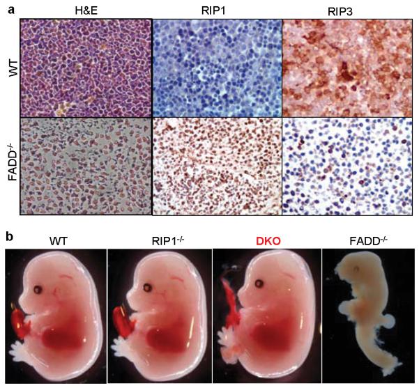
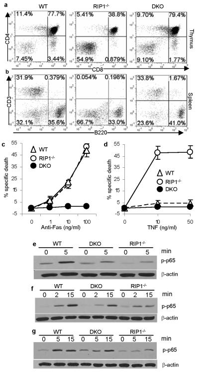
FADD is a common adaptor shared by several death receptors for signalling apoptosis through recruitment and activation of caspase 8 (refs 1-3). Death receptors are essential for immune homeostasis, but dispensable during embryogenesis. Surprisingly, Fadd(-/-) mice die in utero and conditional deletion of FADD leads to impaired lymphocyte proliferation. How FADD regulates embryogenesis and lymphocyte responses has been a long-standing enigma. FADD could directly bind to RIP1 (also known as RIPK1), a serine/threonine kinase that mediates both necrosis and NF-κB activation. Here we show that Fadd(-/-) embryos contain raised levels of RIP1 and exhibit massive necrosis. To investigate a potential in vivo functional interaction between RIP1 and FADD, null alleles of RIP1 were crossed into Fadd(-/-) mice. Notably, RIP1 deficiency allowed normal embryogenesis of Fadd(-/-) mice. Conversely, the developmental defect of Rip1(-/-) lymphocytes was partially corrected by FADD deletion. Furthermore, RIP1 deficiency fully restored normal proliferation in Fadd(-/-) T cells but not in Fadd(-/-) B cells. Fadd(-/-)Rip1(-/-) double-knockout T cells are resistant to death induced by Fas or TNF-α and show reduced NF-κB activity. Therefore, our data demonstrate an unexpected cell-type-specific interplay between FADD and RIP1, which is critical for the regulation of apoptosis and necrosis during embryogenesis and lymphocyte function.
Figures

Comment in
-
Programmed cell death: Apoptosis meets necrosis.Nature. 2011 Mar 17;471(7338):310-2. doi: 10.1038/471310a. Nature. 2011. PMID: 21412328 No abstract available.
-
Cell death: A killer puts a stop on necroptosis.Nat Rev Mol Cell Biol. 2011 May;12(5):279. doi: 10.1038/nrm3101. Epub 2011 Mar 30. Nat Rev Mol Cell Biol. 2011. PMID: 21448226 No abstract available.
Similar articles
-
Programmed cell death: Apoptosis meets necrosis.Nature. 2011 Mar 17;471(7338):310-2. doi: 10.1038/471310a. Nature. 2011. PMID: 21412328 No abstract available.
-
RIP1 kinase activity-dependent roles in embryonic development of Fadd-deficient mice.Cell Death Differ. 2017 Aug;24(8):1459-1469. doi: 10.1038/cdd.2017.78. Epub 2017 Jun 2. Cell Death Differ. 2017. PMID: 28574501 Free PMC article.
-
RIP1-mediated regulation of lymphocyte survival and death responses.Immunol Res. 2011 Dec;51(2-3):227-36. doi: 10.1007/s12026-011-8249-3. Immunol Res. 2011. PMID: 22038529 Free PMC article. Review.
-
The death domain of FADD is essential for embryogenesis, lymphocyte development, and proliferation.J Biol Chem. 2009 Apr 10;284(15):9917-26. doi: 10.1074/jbc.M900249200. Epub 2009 Feb 9. J Biol Chem. 2009. PMID: 19203997 Free PMC article.
-
RIPK-dependent necrosis and its regulation by caspases: a mystery in five acts.Mol Cell. 2011 Oct 7;44(1):9-16. doi: 10.1016/j.molcel.2011.09.003. Mol Cell. 2011. PMID: 21981915 Free PMC article. Review.
Cited by
-
NFκB and ubiquitination: partners in disarming RIPK1-mediated cell death.Immunol Res. 2012 Dec;54(1-3):214-26. doi: 10.1007/s12026-012-8321-7. Immunol Res. 2012. PMID: 22477525 Review.
-
Death receptor-ligand systems in cancer, cell death, and inflammation.Cold Spring Harb Perspect Biol. 2013 May 1;5(5):a008698. doi: 10.1101/cshperspect.a008698. Cold Spring Harb Perspect Biol. 2013. PMID: 23637280 Free PMC article. Review.
-
Border Security: The Role of RIPK3 in Epithelium Homeostasis.Front Cell Dev Biol. 2016 Jun 28;4:70. doi: 10.3389/fcell.2016.00070. eCollection 2016. Front Cell Dev Biol. 2016. PMID: 27446921 Free PMC article.
-
The Tumor Suppressor Hace1 Is a Critical Regulator of TNFR1-Mediated Cell Fate.Cell Rep. 2016 May 17;15(7):1481-1492. doi: 10.1016/j.celrep.2016.04.032. Epub 2016 May 5. Cell Rep. 2016. PMID: 27160902 Free PMC article.
-
RIP3: a molecular switch for necrosis and inflammation.Genes Dev. 2013 Aug 1;27(15):1640-9. doi: 10.1101/gad.223321.113. Genes Dev. 2013. PMID: 23913919 Free PMC article. Review.
References
-
- Boldin MP, et al. A novel protein that interacts with the death domain of Fas/APO1 contains a sequence motif related to the death domain. J. Biol. Chem. 1995;270:7795–7798. - PubMed
-
- Chinnaiyan AM, O’Rourke K, Tewari M, Dixit VM. FADD, a novel death domain-containing protein, interacts with the death domain of Fas and initiates apoptosis. Cell. 1995;81:505–512. - PubMed
-
- Zhang J, Cado D, Chen A, Kabra NH, Winoto A. Absence of Fas-mediated apoptosis and T cell receptor-induced proliferation in FADD-deficient mice. Nature. 1998;392:296–300. - PubMed
-
- Yeh W-C, et al. FADD: essential for embryo development and signaling from some, but not all, inducers of apoptosis. Science. 1998;279:1954–1958. - PubMed
Publication types
MeSH terms
Substances
Grants and funding
- AI083497/AI/NIAID NIH HHS/United States
- AI017672/AI/NIAID NIH HHS/United States
- DK32520/DK/NIDDK NIH HHS/United States
- R01 AI083497/AI/NIAID NIH HHS/United States
- AI076788/AI/NIAID NIH HHS/United States
- CA95454/CA/NCI NIH HHS/United States
- R03 AI076788/AI/NIAID NIH HHS/United States
- AI083915/AI/NIAID NIH HHS/United States
- R01 AI017672/AI/NIAID NIH HHS/United States
- R01 CA095454/CA/NCI NIH HHS/United States
- P30 DK032520/DK/NIDDK NIH HHS/United States
- R21 AI083915/AI/NIAID NIH HHS/United States
- R37 AI017672/AI/NIAID NIH HHS/United States
LinkOut - more resources
Full Text Sources
Other Literature Sources
Molecular Biology Databases
Research Materials
Miscellaneous
